Diffusion imaging differs from traditional MR technology as it primarily relies on the movement of water molecules rather than the spin proton density of tissues, T1 values, or T2 values, providing a novel technique for tissue imaging contrast. The diffusion of water molecules in tissues is a random thermal motion, where the direction and magnitude of diffusion are influenced by the local microenvironment of the tissue. For instance, in areas affected by cerebral infarction or tumors, the diffusion of water molecules is restricted, leading to a decrease in diffusivity.
With the advancement of nuclear magnetic technology, various diffusion models have been proposed, such as Intra-Voxel Incoherent Motion (IVIM), Diffusion Kurtosis Imaging (DKI), Diffusion Tensor Imaging (DTI), Diffusion Spectrum Imaging (DSI), High Angular Resolution Diffusion Imaging (HARDDI), and Neurite Orientation Dispersion and Density Imaging (NODDI).
Increasingly complex models require more b values and more gradient directions. However, as the b values and gradient directions increase, the scan time also increases. In this case, studies of white matter fiber bundles typically use conventional DTI scans, often employing relatively large voxels (e.g., 3x3x3 mm3), a single b value (b = 1000 s/mm2), and a limited number of gradient directions (32 directions). The Human Connectome Project (HCP) uses conventional diffusion scanning parameters on the 3T magnetic resonance MAGNETOM Skyra: 1.25×1.25×1.25 mm3, three b values (b = 1000, 2000, 3000 s/mm2), and 270 gradient directions. Such parameters undoubtedly improve imaging quality significantly, but the time required also increases. The project team innovatively combined the latest SMS diffusion EPI technology with an acceleration factor of 3 (Ugurbil et al., 2013), achieving a balance between quality and speed.
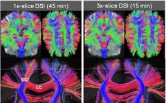
Using simultaneous multi-slice imaging technology can significantly reduce scan time. For instance, acquiring a voxel of 2.5×2.5×2.5 mm3, with a maximum b value of 7000 s/mm2, 256 directions, and acquiring a B0 image every 20 TR increases the DSI. If conventional diffusion EPI sequences are used, scanning the entire brain takes 45 minutes, whereas using the SMS diffusion EPI sequence with an acceleration factor of 3 can reduce the scan time to 15 minutes, with nearly no impact on image signal-to-noise ratio (Feinberg and Setsompop, 2013).
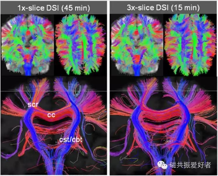
Figure 1, DSI images obtained from Siemens 3T magnetic resonance (Feinberg and Setsompop, 2013).
Simultaneous multi-slice diffusion imaging technology can be applied not only to the head but also to the scanning of the liver, kidneys, and pelvis, and can perform large-area diffusion imaging. A study from New York University showed that using the SMS diffusion EPI sequence for diffusion-weighted imaging of the liver on a Siemens 3T magnetic resonance MAGNETOM Skyra reduced the scan time by 40% compared to traditional diffusion sequences, with no change in image signal-to-noise ratio (Obele et al., 2015).
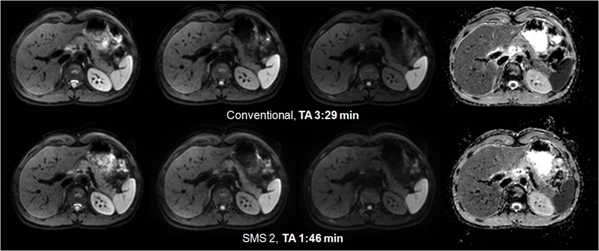 Figure 2. Simultaneous multi-slice abdominal diffusion imaging
Conventional diffusion imaging sequences use single-shot EPI for image acquisition, which can easily produce phase errors and geometric distortions in areas of inhomogeneous magnetic fields, such as air-tissue interfaces and muscle-bone interfaces. Additionally, due to the need for a certain diffusion time, the TE is relatively long, and using high spatial resolution acquisition can lead to a decrease in SNR. To address this issue, Siemens introduced the readout segmented EPI sequence called RESOLVE. This sequence has been promoted for use in Siemens’ next-generation magnetic resonance products. Using RESOLVE can significantly improve image signal-to-noise ratio, reduce geometric distortion, and enhance spatial resolution. It has significant clinical application value for detecting small cerebral infarcts, spinal cord injuries, and tumor localization. By employing simultaneous multi-slice imaging technology, RESOLVE can be further optimized to increase its scanning speed.
Siemens magnetic resonance engineer Ms. Liu Wei demonstrated the head and spinal cord large-area DTI images obtained using the SMS RESOLVE sequence on the Siemens 3T magnetic resonance at ISMRM in 2015. The spatial resolution was 2.5×2.5×2.5 mm3, with a b value of 800 s/mm2, gradient directions of 30, and segmentation of 5. If the normal RESOLVE sequence is used, the scan time can reach 25 minutes, which is unacceptable for general clinical examinations and research. When the acceleration factor is 2, the scan time is shortened to 13 minutes; when the acceleration factor is 3, the scan time is reduced to under 9 minutes (Figure 3). This same technology can also be applied to breast diffusion-weighted imaging.
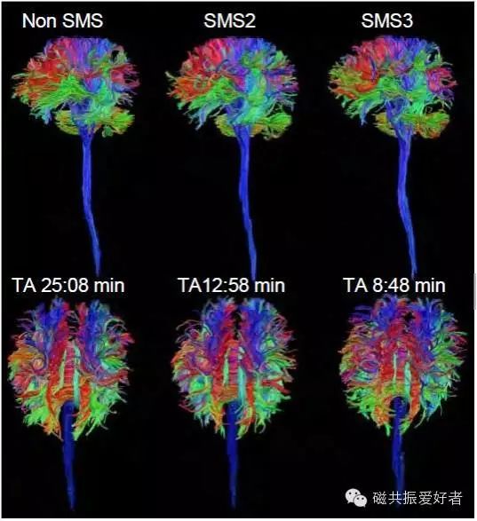
Figure 3. Large-area DTI images of the head and spine obtained using SMS RESOLVE (Wei Liu et al., 2015, ISMRM).
As scan times increase, patient cooperation significantly decreases, making it impossible to complete the examination; or if completed reluctantly, numerous motion-related artifacts may arise, affecting diagnostic results. Previously, advanced complex diffusion models were often used for research rather than routine clinical use. With the introduction of SMS, the bottlenecks faced by diffusion imaging have been resolved, allowing advanced diffusion imaging methods to be easily integrated into routine clinical practice.
In the next issue, I will bring you the progress of SMS applications in functional imaging, so stay tuned.
References
Feinberg, D.A., and Setsompop, K. (2013). Ultra-fast MRI of the human brain with simultaneous multi-slice imaging. J Magn Reson 229, 90-100.
Obele, C.C., Glielmi, C., Ream, J., Doshi, A., Campbell, N., Zhang, H.C., Babb, J., Bhat, H., and Chandarana, H. (2015). Simultaneous Multislice Accelerated Free-Breathing Diffusion-Weighted Imaging of the Liver at 3T. Abdom Imaging.
Ugurbil, K., Xu, J., Auerbach, E.J., Moeller, S., Vu, A.T., Duarte-Carvajalino, J.M., Lenglet, C., Wu, X., Schmitter, S., Van de Moortele, P.F., et al. (2013). Pushing spatial and temporal resolution for functional and diffusion MRI in the Human Connectome Project. NeuroImage 80, 80-104.
Click the button in the upper right corner to share with your friends and let more people know about the latest developments in magnetic resonance.
Scan the QR code below to follow 【Magnetic Resonance Enthusiasts】