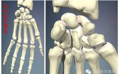
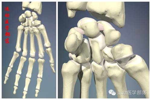
Joint Structure
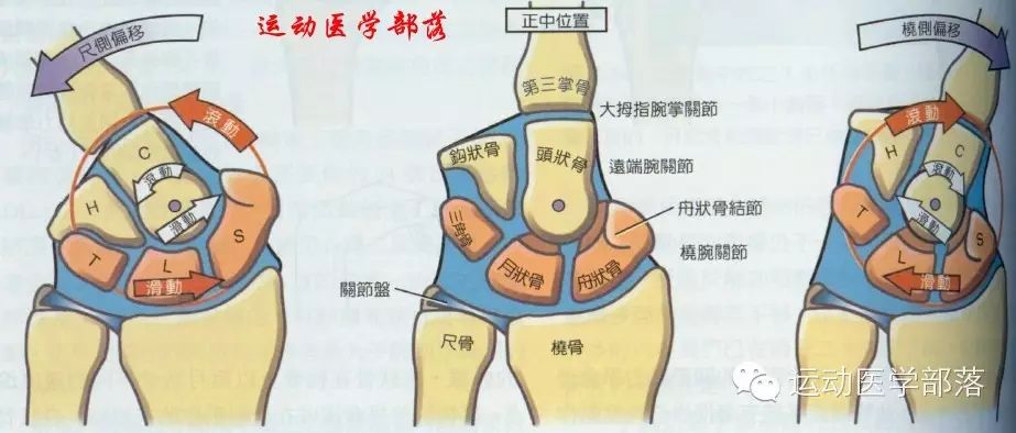
The wrist joint consists of the radiocarpal joint and intercarpal joints. The radiocarpal joint is a condyloid joint formed by the concave distal end of the radius and the articular disc, which articulates with the convex surfaces of the scaphoid, lunate, and triquetrum bones. Some scholars do not consider the ulnar and pisiform bones as part of this joint.
The hand consists of the carpometacarpal joints (CMC), metacarpophalangeal joints (MCP), and interphalangeal joints (PIP, DIP).
Kinematics of the Wrist & Hand
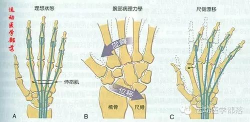
The convex proximal row of carpal bones moves in the opposite direction of hand movement. During wrist flexion, the carpal bones slide backward over the radius and articular disc; during wrist extension, they slide forward; during radial deviation, they slide toward the ulnar side; and during ulnar deviation, they slide toward the radial side.
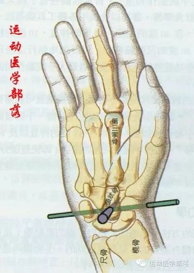
The axis of rotation for the wrist joint is considered to pass through the head of the capitate bone, and this axis shifts inward and outward with flexion and extension, and shifts anteriorly and posteriorly with radial and ulnar deviation.
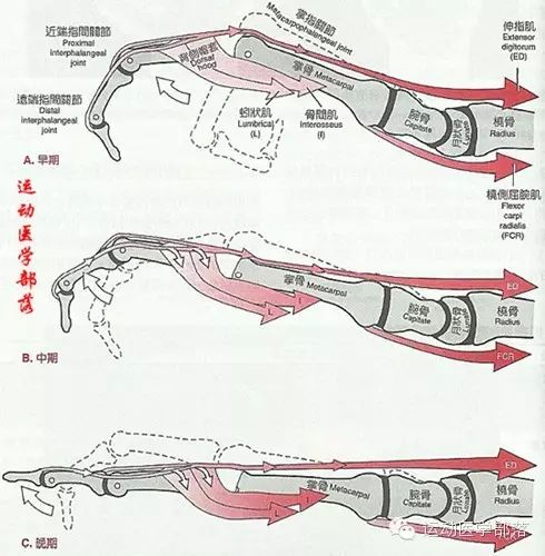
The metacarpophalangeal joints (MCP) of the fingers are biaxial joints. The convex and rounded heads of the metacarpals articulate with the concave bases of the proximal phalanges, allowing movements such as flexion, extension, and hyperextension, along with abduction and adduction.
The finger joints are uniaxial hinge joints, allowing only flexion and extension movements.
Summary of Muscle Innervations
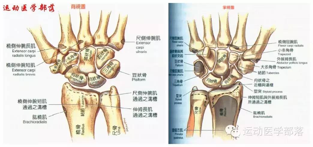
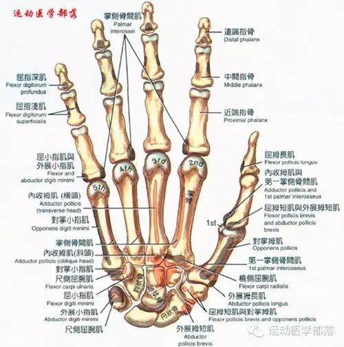
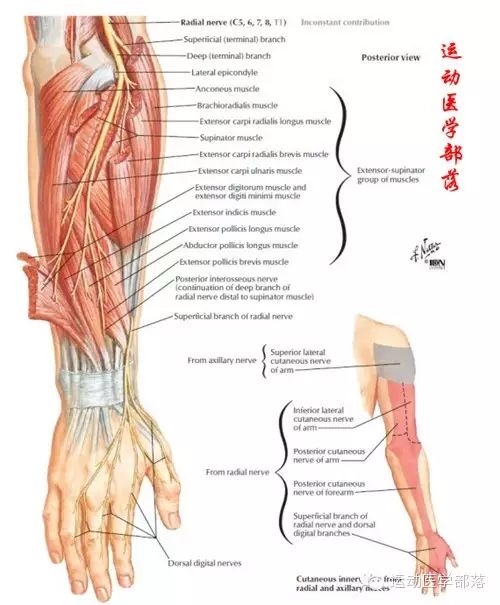
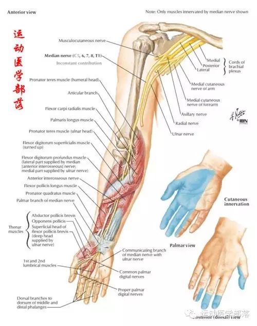
The radial nerve innervates the muscles on the posterior side; the median nerve innervates the muscles on the anterior side of the thumb; and the ulnar nerve innervates the muscles on the ulnar side.
The interaction between agonist and antagonist muscles during wrist and hand movements:
The interaction between intrinsic and extrinsic muscles of the fingers:

Stability
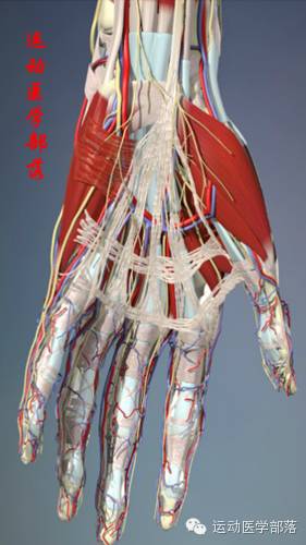
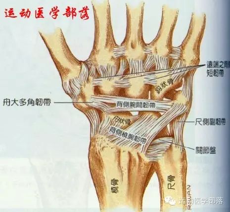
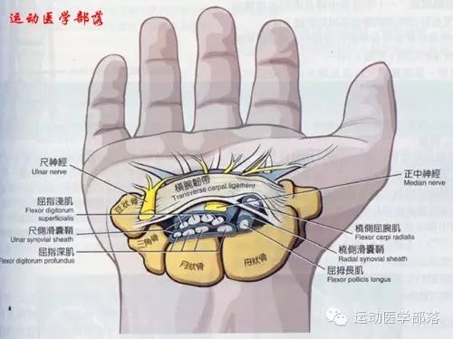
Factors contributing to the stability of the wrist and hand joints include: bone configuration, ligament tension, and muscle support. Here, I briefly introduce some commonly used ligaments: radial collateral ligament, palmaris longus tendon, dorsal radiocarpal ligament, palmar fascia, flexor retinaculum, palmar carpal ligament, transverse carpal ligament, extensor retinaculum, extensor expansion ligament, as well as proximal and distal carpal arches, and longitudinal arches.
Common Injury at the Wrist
Carpal tunnel syndrome: the median nerve is compressed within the carpal tunnel at the wrist.
Wrist instability: ulnar-sided “variant”; instability of the metacarpophalangeal joints, etc.
Joint deformities caused by rheumatoid arthritis: swan-neck deformity.
Avascular necrosis of the lunate bone.
Palmar aponeurosis contracture.
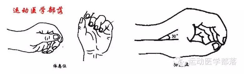
Resting position of the hand: Wrist joint extension 10-15 degrees, with slight ulnar deviation, thumb slightly abducted, thumb tip touching the radial side of the distal interphalangeal joint of the index finger, fingers from index to little finger in a half-flexed position, index finger slightly flexed, little finger more flexed, index finger slightly ulnar deviated, little finger slightly radially deviated. In this position, the flexor tendons are in a balanced state; if the hand is injured, this balance is disrupted.
Functional position of the hand: Wrist joint extension 30 degrees, with about 10 degrees of ulnar deviation, metacarpophalangeal joints flexed 30-45 degrees, proximal interphalangeal joints flexed 60-80 degrees, distal interphalangeal joints slightly flexed about 10-15 degrees. Fingers spread apart, thumb in abduction relative to the palm. In the functional position, the hand can perform its maximum function. Therefore, after a hand injury, fractures usually require the hand to be fixed in the functional position.
(Reference: Fundamentals of Kinesiology and Rehabilitation Medicine, NTUPT, some images sourced from the internet.)