It is believed that when patients undergo cataract surgery on the contralateral eye, they are often told that “the second eye is generally more sensitive than the first eye.” But why is this? Previous studies suggested that it might be related to psychological factors, such as the patient’s preoperative tension and anxiety being lower for the second eye than for the first. Recently, an article published in IOVS provided a possible new explanation from the perspective of cytokines.
The researchers from Fudan Eye, Ear, Nose and Throat Hospital believe that the increased sensitivity of the contralateral eye may be related to sympathetic responses. It is well known that pain is associated with inflammation, and many inflammatory cytokines are related to pain, such as IL-1, IL-6, IL-8, MIP (Macrophage Inflammatory Protein), MCP-1 (Monocyte Chemoattractant Protein 1), and RANTES (Regulated Upon Activation, Normal T Cell Expressed and Secreted). Thus, the hypothesis was proposed: Is the increased sensitivity of the contralateral eye to pain related to cytokines?
Methods: The experimental group included 51 patients who underwent cataract surgery on their second eye, all of whom had surgery on the first eye one month prior, using PHACO with no other ocular complications. The control group included 50 patients undergoing cataract surgery on their first eye. The specimen collection method involved using a 26G needle to perform anterior chamber paracentesis at the start of the surgery, collecting 100-200μL of aqueous humor. A protein antibody chip (RayBiotech) was used to simultaneously detect 40 cytokines in the aqueous humor. Based on the preliminary screening results, one cytokine (IL-1ra: IL-1 receptor antagonist) and four chemokines (MCP-1, MIP-1a, MIP-1b, RANTES) were selected for further concentration detection using Bio-Plex suspension cytokine chip technology.
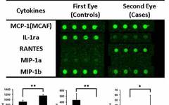
Results: The initial screening of 40 cytokines showed that the expression of IL-1ra, MIP-1a, and MIP-1b in the control group was significantly higher than that in the experimental group, while MCP-1 and RANTES were the opposite, with significantly higher expression in the experimental group (P<0.05) (Figure 1). However, after Bonferroni correction, only MCP-1 and IL-1ra showed statistical significance (P<0.00125). Further detection of the concentrations of five cytokines using suspension cytokine chip technology indicated that only MCP-1 had statistical significance between the two groups (P<0.05).
Discussion and Conclusion: This study compared the expression and concentration of cytokines in the aqueous humor of the first and second eyes during cataract surgery and found that MCP-1 was elevated in the second eye. Many cells can produce MCP-1, especially monocytes and macrophages, which can chemotactically aggregate leukocytes during inflammation and tissue damage. Increased expression of MCP-1 is also observed in many inflammation-related diseases, such as atherosclerosis and arthritis. MCP-1 is also a pain-related factor, and studies have shown that the expression level of MCP-1 is closely related to the degree of pain in fibromyalgia. The increased concentration of MCP-1 in the aqueous humor of the contralateral eye may be the pathophysiological basis for the increased sensitivity during surgery. As for the reason for its elevation, it is speculated that it may be caused by a sympathetic response similar to sympathetic ophthalmia. It is currently believed that T helper cell-mediated delayed-type hypersensitivity is the main mechanism of sympathetic ophthalmia, and many cytokines can mediate its downstream responses, including MCP-1. Of course, the results and conclusions of this study need further research support.
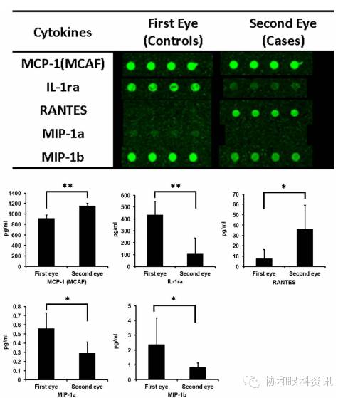
Figure 1: Results of the initial screening using a chip for detecting 40 cytokines, where differences were found in five cytokines between the two groups.
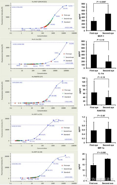
Figure 2: Further detection of five cytokines using suspension cytokine chip technology, showing significant differences in expression for MCP-1, which was higher in the experimental group compared to the control group.
(All images in the text are extracted from the original article)
Original Article: Zhu X-J, Wolff D, Zhang K-K, et al. Molecular inflammation in the contralateral eye after cataract surgery in the first eye. Invest Ophthalmol Vis Sci. 2015;56:5566–5573. DOI:10.1167/iovs.15-16531
Comment: It is not uncommon in clinical practice for patients to report more significant pain during the second eye cataract surgery compared to the first eye. The authors of this article were able to propose a reasonable hypothesis based on the pathogenesis of sympathetic ophthalmia and validate it through basic research, reflecting a translational medicine approach from bedside to bench that we should learn from. The authors confirmed the differences in MCP-1 between the second eye and the control eye using two high-throughput protein detection technologies, and the results are quite credible. However, this article also exposes some limitations of the current trace protein detection methods for aqueous humor: for the various cytokines mentioned in the text, the differences in concentrations measured between the second eye group and the control group were inconsistent between the two methods, such as MIP-1a and MIP-1b, where the first method showed higher values in the control group compared to the second eye, while the second method showed the opposite, indicating that the methodology of high-throughput detection of aqueous humor cytokines still needs improvement. Particularly for the second method, only the concentration range of MCP-1 was within the linear range of the standard curve, while the concentrations of the other cytokines were below the linear range, thus the reliability of the detection values is not high.
—— Zhao Chan
Source: Xiehe Eye News