Key Points in the Diagnosis and Treatment of Metacarpophalangeal Joint Dislocation

Click the aboveGood Medicine, follow us

Key Points in the Diagnosis and Treatment of Metacarpophalangeal Joint Dislocation

Introduction

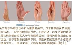
The metacarpophalangeal joint primarily performs flexion and extension, with a few individuals able to hyperextend. The degree of flexion varies significantly. The metacarpophalangeal joint often dislocates due to external forces caused by finger sprains, punctures, or extreme dorsal extension or flexion. Depending on the position of the finger during trauma and the direction of the external force, the metacarpophalangeal joint can experience dorsal or palmar dislocation, with dorsal dislocation being more common. Recently, during my training in the Trauma Orthopedics department of Weihai Municipal Hospital, I encountered two cases of thumb MCP joint dislocation (one palmar and one dorsal dislocation). Throughout the management of these cases, I gained deeper insights into the treatment of metacarpophalangeal joint dislocations. I would like to thank my mentors, Associate Chief Physician Xiao Hongqiang, Associate Chief Physician Chen Kang, attending physician Li Dawei, and other teachers in the Trauma Orthopedics department for their guidance and support during my orthopedic training. This article aims to present relevant knowledge on thumb metacarpophalangeal joint dislocation through the collection and organization of related literature, hoping to enhance understanding of metacarpophalangeal joint dislocations, especially those of the thumb, and to facilitate clinical management of such conditions.
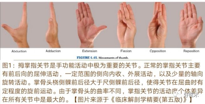
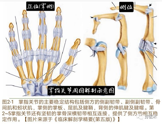
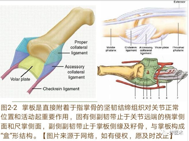
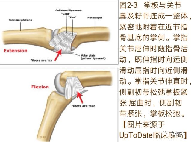
Key Points in the Diagnosis and Treatment of Metacarpophalangeal Joint Dislocation

1. Anatomy

The main stabilizing structures of the metacarpophalangeal joint include the collateral ligaments, accessory collateral ligaments, interosseous muscles, and lumbrical muscles on the lateral side; the palmar plate, flexor muscles, and tendon sheaths on the palmar side; and the extensor tendons and tendon hood on the dorsal side. The second to fifth metacarpophalangeal joints are interconnected by the strong deep transverse metacarpal ligament, providing lateral stability (Figure 2-1).

Key Points in the Diagnosis and Treatment of Metacarpophalangeal Joint Dislocation

The palmar plate, also known as the palmar ligament (Figure 2-2), is a rectangular dense fibrocartilaginous plate located on the palmar side of the metacarpophalangeal joint. Its distal end is thick and robust, attaching to the palmar lip of the base of the proximal phalanx, while the proximal end is thin and lax, forming a membrane-like attachment to the palmar side of the metacarpal neck and intertwining with the deep fascia, connecting laterally with the collateral ligaments. The origin of the palmar plate is shaped like a swallowtail, connected to the phalanx and fibrous tendon sheath only by the proximal checkrein ligament. The distal end of the palmar plate gradually becomes cartilaginous, with its central part attaching to the periosteum and the lateral part attaching to the collateral ligaments.

Key Points in the Diagnosis and Treatment of Metacarpophalangeal Joint Dislocation

The collateral ligaments originate from the dorsal side of the joint and attach to the palmar side of the joint. The proper collateral ligaments attach to the radial palmar side and the ulnar palmar side of the distal joint, while the accessory collateral ligaments attach to the lateral edge of the palmar plate and the sesamoid bones, forming a “box” structure (Figure 2-3).

Key Points in the Diagnosis and Treatment of Metacarpophalangeal Joint Dislocation

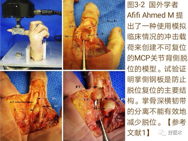
2. Causes and Mechanisms
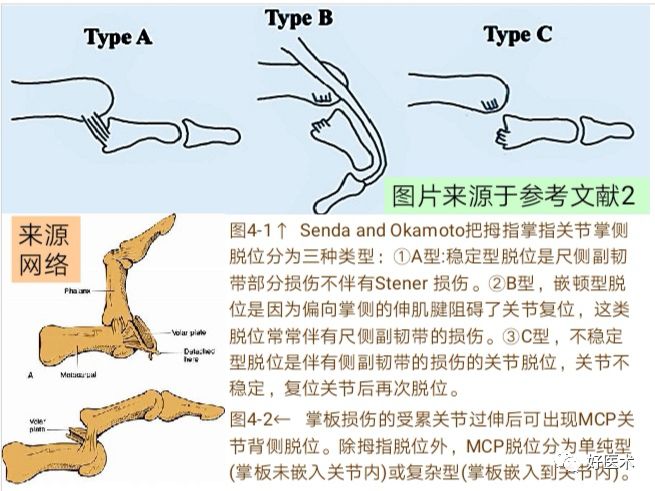
MCP joint dislocation is a rare condition. Except for the thumb, the metacarpophalangeal joints are protected by their anatomical position and the strong ligamentous complex, making dislocation uncommon. The position of the thumb itself, as well as the surrounding positions of the index and little fingers, makes them more susceptible to such injuries.
Dorsal dislocation of the metacarpophalangeal joint primarily occurs due to indirect forces causing finger sprains, punctures, or extreme dorsal extension. Overextension injuries to the metacarpophalangeal joint can lead to complete dislocation, with the torn palmar plate (sometimes along with the sesamoid bone) becoming trapped in the joint, hindering reduction.

Key Points in the Diagnosis and Treatment of Metacarpophalangeal Joint Dislocation

Dorsal subluxation often occurs due to excessive hyperextension forces acting on the thumb metacarpal, often resulting in a tear of the proximal palmar plate. Dorsal subluxation of the thumb metacarpophalangeal joint is also known as simple dorsal dislocation, where the surfaces between the metacarpal and phalanx still maintain contact, but most joint connections remain intact.

Key Points in the Diagnosis and Treatment of Metacarpophalangeal Joint Dislocation

Palmar dislocation of the metacarpophalangeal joint is a rare traumatic injury. The recognized mechanism of injury for this condition is extreme flexion of the metacarpophalangeal joint, leading to dorsal capsular damage, tendon injury, and rupture of the medial collateral ligament, resulting in palmar dislocation of the joint. Wang Guan et al. proposed in their case report on palmar dislocation of the metacarpophalangeal joint that the likely cause of palmar dislocation is: when the interphalangeal joint of the thumb is straightened, the metacarpophalangeal joint is excessively flexed, and the thumb is in an abducted position, vertical pressure from the tip of the thumb to the metacarpophalangeal joint is applied.

3. Classification

Metacarpophalangeal joint dislocations can be classified into dorsal and palmar dislocations, with dorsal dislocations being the most common. Additionally, there are lateral dislocations, which mainly involve collateral ligament injuries and will not be discussed here.
Dorsal dislocation occurs when the proximal phalanx gets stuck on the dorsal radial side of the metacarpal, often presenting with an angle of 60°-90° between the phalanx and metacarpal, with joint surfaces still in contact. Closed reduction should be performed.
Note:Dislocations can be classified as “simple” or “complex”. Simple dislocations present with a certain angle between the proximal phalanx and the metacarpal axis, and subluxations can sometimes appear similarly. In simple dislocations, closed reduction can be performed after adequate anesthesia; complex dislocations occur when the proximal phalanx overlaps with the metacarpal (the axes of both are parallel).In complex dislocations, the palmar plate becomes trapped in the joint space, and its presentation resembles that of simple dislocation, but the angle is usually smaller.The proximal phalanx often runs parallel to the metacarpal.Soft tissue trapped in the joint space complicates closed reduction, often making it impossible and usually requiring surgery.Closed reduction should be attempted first.
Palmar dislocations are rare, requiring greater force to cause the injury, often resulting in severe damage to the joint capsule and collateral ligaments.Surgical treatment is usually necessary, but closed reduction should be attempted first.

Key Points in the Diagnosis and Treatment of Metacarpophalangeal Joint Dislocation

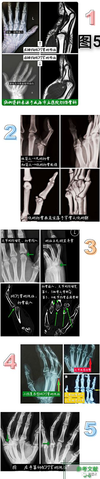
4. Clinical Diagnosis
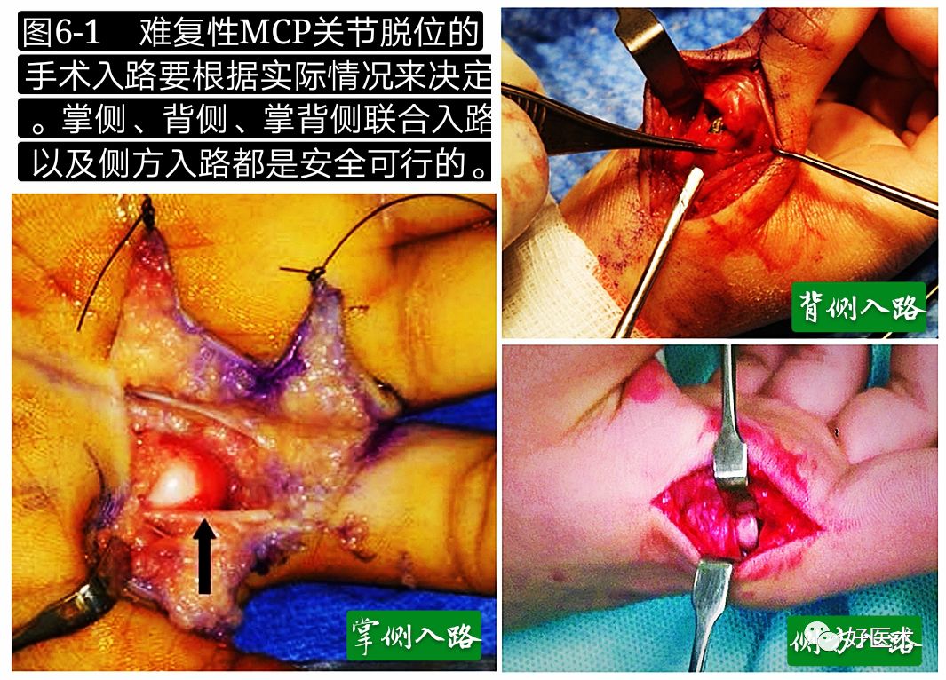
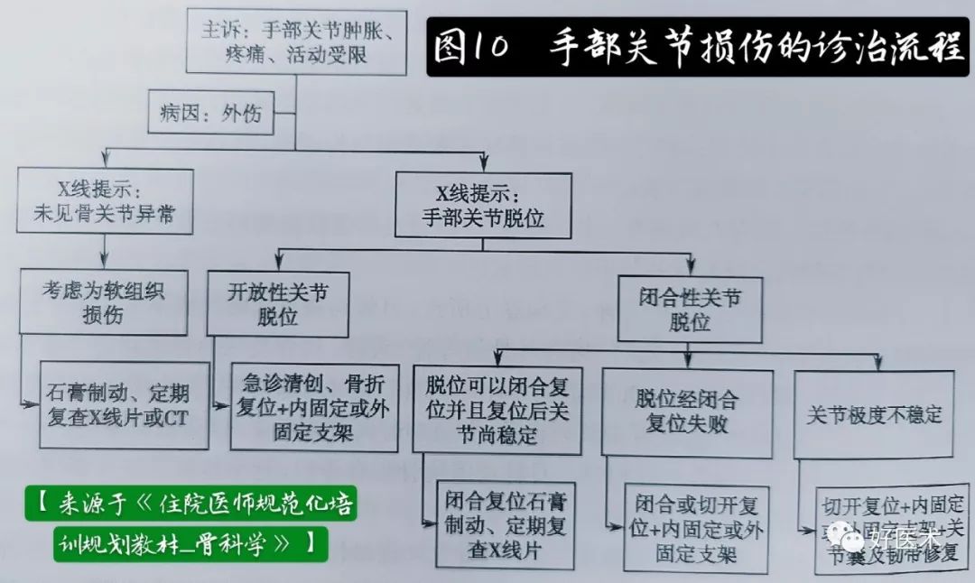
Joint dislocation is a rare injury, with the index finger being the most commonly affected, followed by the thumb. After dorsal dislocation of the metacarpophalangeal joint, the phalanx shifts dorsally, and the metacarpal head protrudes towards the palmar side, forming a deformity of joint hyperextension, which can manifest as local swelling, pain, and functional impairment.
Diagnosis of metacarpophalangeal joint dislocation is not difficult. Clinically, there will be significant hyperextension deformity of the metacarpophalangeal joint, with the metacarpal in an adducted position. If there is a depression in the skin over the palmar side of the metacarpophalangeal joint, it indicates a complex dislocation.
X-ray films:including anteroposterior, lateral, and oblique views, are the initial assessments for thumb injuries. The lateral view should be a true lateral view of the thumb (i.e., the bones of the thumb arranged appropriately without rotation), not the lateral view of the hand. When assessing metacarpophalangeal joints that are difficult to reduce or cannot be reduced, ① if the gap between the metacarpal head and the proximal phalanx increases, it indicates the possibility of soft tissue entrapment. ② If a sesamoid bone is trapped between the metacarpal head and the proximal phalanx, it suggests a complex irreducible dislocation.
CT and 3D reconstructioncan further clarify the type of dislocation, changes in the joint space, and alterations in bony structures. MRI examinations provide a more accurate assessment of the degree of soft tissue damage around the metacarpophalangeal joint, especially the collateral ligaments.

Key Points in the Diagnosis and Treatment of Metacarpophalangeal Joint Dislocation

5. Treatment

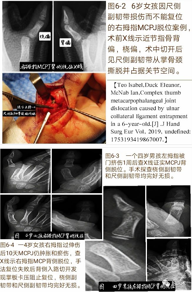
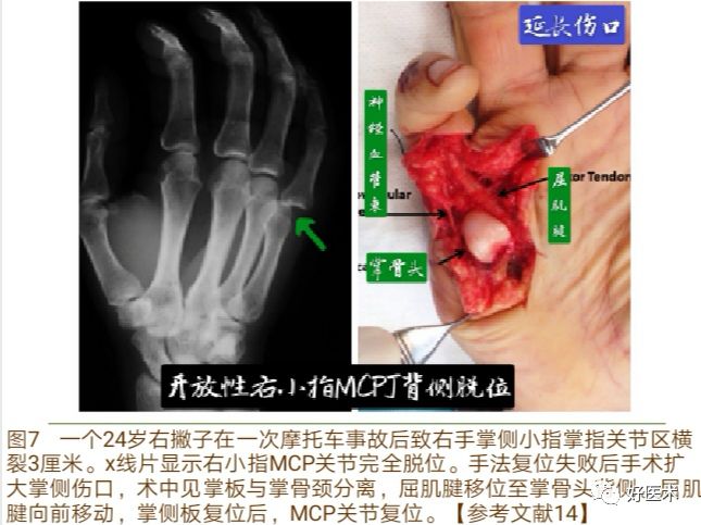
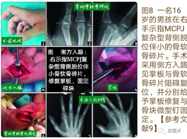
1. Treatment of Dorsal Dislocation

Most dorsal dislocations can be easily reduced without surgery. Manual reduction should avoid simple traction on the thumb, as this can turn a simple dislocation into a complex irreducible dislocation. The recommended reduction method is to apply pressure to the proximal phalanx while extending the metacarpophalangeal joint.Longitudinal traction or increasing hyperextension deformity may further displace the palmar plate dorsally, trapping it between the metacarpal head and the base of the proximal phalanx, thus rendering reducible MCP joint subluxation irreducible.After successful reduction, plaster immobilization of the metacarpophalangeal joint in approximately 60° flexion should be performed for 2-3 weeks.
Complex dislocations are difficult to achieve closed reduction because the palmar plate moves dorsally with the phalanx and is tightly pressed against the dorsal side of the metacarpal head, hindering the return of the base of the proximal phalanx to its original position. Nevertheless, complex dislocations should still first attempt closed reduction; only if closed reduction fails or the joint remains unstable after reduction should open reduction be considered.
Choice of Surgical Approach
The surgical approach for irreducible MCP joint dislocations should be determined based on the actual situation. For experienced surgeons, palmar, dorsal, combined palmar-dorsal, and lateral approaches are all safe and feasible. The key is to achieve the desired effect under the safest conditions. Each method has its advantages and disadvantages, and there is still debate about which method is superior.

Key Points in the Diagnosis and Treatment of Metacarpophalangeal Joint Dislocation

Dorsal Approach:(Figure 6-2/3) A slight curved longitudinal incision is made on the dorsal side to expose the extensor tendons, entering between the long and short extensor tendons, exposing the joint capsule.A longitudinal incision is made in the joint capsule.This approach allows for easy exposure of the joint and examination of the proximally subluxated phalanx.It is often possible to find the palmar plate dragged to the dorsal side, covering the metacarpal head.Using a blunt instrument, such as a retractor, to push towards the palmar side often allows for easy release of the palmar plate from the joint.If simple direct pushing is unsuccessful, a longitudinal incision in the palmar plate can make extraction easier.For fixation of associated osteochondral fractures of the metacarpal head, the dorsal approach provides excellent exposure.The advantage is that it allows for pushing the entrapped soft tissue out of the joint towards the palmar side, which is easier than pulling the soft tissue out of the joint from the palmar side. However, once the joint is reduced, the damaged soft tissue on the palmar side of the joint will not be visible.

Key Points in the Diagnosis and Treatment of Metacarpophalangeal Joint Dislocation

Palmar Approach:This approach allows for the exposure of both intrinsic and extrinsic flexors of the joint.As mentioned earlier, these structures often obstruct reduction.After hyperextending the proximal phalanx, the entrapped soft tissue within the joint can be freed.Once the joint is reduced, it is usually stable; however, if the joint is unstable, direct repair or fixation using suture anchors may be required for the collateral ligaments.The advantage is that it allows visualization of the entrapped soft tissue within the joint, which may be the palmar plate or sesamoid bone, and allows for its removal from the base of the metacarpal head and proximal phalanx.If these tissues on the palmar side need repair, it can be performed after joint reduction.(Figure 7)

Key Points in the Diagnosis and Treatment of Metacarpophalangeal Joint Dislocation

Lateral Approach (Figure 8):This approach allows for visualization and management of both palmar and dorsal structures, reduction of the palmar plate, and fixation of osteochondral fragments on the palmar side.Additionally, surgical scarring can reduce the risk of tendon adhesions and minimize scar contraction, a common complication that restricts joint movement.

Key Points in the Diagnosis and Treatment of Metacarpophalangeal Joint Dislocation

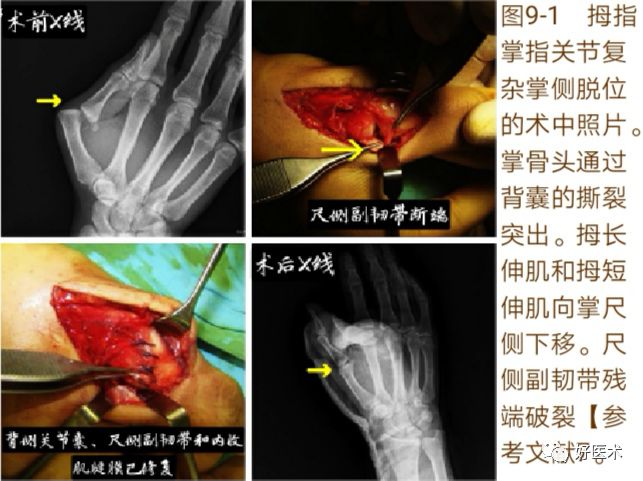
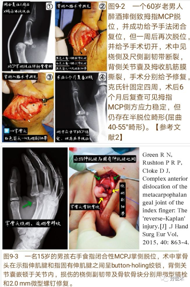
2. Treatment of Palmar Dislocation

Singhal first reported a case of palmar MCP dislocation in 1974, where the vast majority cannot be reduced through closed methods and usually require open surgical reduction (Figure 9).

Key Points in the Diagnosis and Treatment of Metacarpophalangeal Joint Dislocation

Closed reduction techniques include axial traction, followed by slight flexion of the PIPJ, and applying pressure dorsally to the base of the dislocated proximal phalanx. If attempts at closed reduction fail, open reduction must be performed.Structures that may affect closed reduction include the palmar plate, dorsal joint capsule, collateral ligaments, or dislocated extensor tendons.After successful manual closed reduction, the stability of the MCPJ should be assessed. If the joint is unstable, a protective early mobilization protocol may be initiated. If the joint remains unstable, surgical repair may be required.Literature reports indicate that in cases where closed reduction is successful, the stability of the metacarpophalangeal joint is often poor, frequently associated with collateral ligament injuries, necessitating primary surgical treatment.

Key Points in the Diagnosis and Treatment of Metacarpophalangeal Joint Dislocation

Wang Guan et al. mentioned in their article “Open Reduction and Internal Fixation for a Case of Palmar Dislocation of the Thumb Metacarpophalangeal Joint” that the incidence of collateral ligament injuries, especially ulnar collateral ligament injuries, in patients with palmar dislocation of the metacarpophalangeal joint is as high as 69.23%. For patients with palmar dislocation of the metacarpophalangeal joint, due to joint swelling, the false negative rate of lateral stress tests is high. Early open reduction and thorough exploration of the collateral ligaments, joint capsule, and tendons, along with primary repair of the damaged areas, are crucial. Early diagnosis and treatment provide satisfactory functional outcomes for palmar dislocations of the MCP joint.

6. Complications

Many complications in patients with MCP joint dislocation are related to diagnostic failure and inadequate treatment.
① Repeated attempts at closed reduction or prolonged dislocation may lead to post-traumatic arthritis or avascular necrosis of the metacarpal head.
② Joint stiffness is a major complication, which may result from tissue damage during injury or excessive fibrosis due to prolonged immobilization, leading to reduced joint range of motion. Additionally, delayed treatment may lead to worse outcomes. Arthrolysis or tendon release may be necessary when needed.
③ Neurovascular bundle injury may occur during open reduction surgery.
④ Premature closure of the growth plate is a rare sequela of MCP joint dislocation in children.
Note: In children whose skeletal development is not yet mature, the growth plate of the phalanx is more fragile than the supporting ligaments. Therefore, Salter-Harris fractures of the phalanx are more common than dislocations. X-rays should first be used to rule out the occurrence of avulsion fractures.

Key Points in the Diagnosis and Treatment of Metacarpophalangeal Joint Dislocation

References
1. Afifi Ahmed M, Medoro Amanda, Salas Christina et al. A cadaver model that investigates irreducible metacarpophalangeal joint dislocation.[J] .J Hand Surg Am, 2009, 34: 1506-11.
2. H Senda, HO Kamoto. Palmar dislocation of the thumb metacarpophalangeal joint: report of four cases and a review of the literature. The Journal of Hand Surgery (European Volume) 2014, 39E(3):276-281.
3. Xiong G, Gao Y, Guo S et al. Pathoanatomy and treatment modifications of metacarpophalangeal joint locking of the thumb.[J] .J Hand Surg Eur Vol, 2015, 40: 68-75.
4. Chitnis Shreyas Sanjeev, Chitnis Sanjeev Lakshaman, Indication for CT following index finger metacarpophalangeal joint dislocation – A case report.[J] .Trauma Case Rep, 2018, 18: 31-36.
5. Ramzi Z, Chafik R, Madhar M et al. Volar metacarpophalangeal joint dislocation: A case report.[J] .Hand Surg Rehabil, 2018, undefined: undefined.
6. Elghoul N, Bouya A, Jalal Y, Zaddoug O, Benchakroun M, Jaafar A. Complex metacarpophalangeal joint dislocation of the little finger: A sesamoid bone seen within joint. What does it mean?. Trauma Case Rep. 2019;23:100225.
7. Yüksel, Serdar & Adanır, Oktay & Beytemür, Ozan & Akif Gülec, Mehmet. (2017). Volar dislocation of the metacarpophalangeal joint of the thumb: A case report. Acta Orthopaedica et Traumatologica Turcica. 51. 10.1016/j.aott.2017.03.014.
8. Teo Isabel, Duck Eleanor, McNab Ian, Complex thumb metacarpophalangeal joint dislocation caused by ulnar collateral ligament entrapment in a 6-year-old.[J] .J Hand Surg Eur Vol, 2019, undefined: 1753193419867007.
9. Pereira JM, Quesado M, Silva M, Carvalho JDD, Nogueira H, Alves J. The Lateral Approach in the Surgical Treatment of a Complex Dorsal Metacarpophalangeal Joint Dislocation of the Index Finger. Case Rep Orthop. 2019;2019:1063829.
10. Chen Xiaodong, Wang Yuren, Yao Weifang. Diagnosis and treatment of thumb simple palmar plate injury leading to metacarpophalangeal joint lock [J]. Chinese Journal of Hand Surgery, 2006, 2:95-96.
11. Guo Wenlai. Case report and literature review of four cases of palmar dislocation of the thumb metacarpophalangeal joint [D]. Changchun: Jilin University, 2015.
12. Green R N, Rushton P R P, Cloke D J, Complex anterior dislocation of the metacarpophalangeal joint of the index finger: The ‘reverse-Kaplan’ injury.[J] .J Hand Surg Eur Vol, 2015, 40: 863-4.
13. Wang Guan, Li Rui, Li Bingwan et al. Open reduction and internal fixation for a case of palmar dislocation of the thumb metacarpophalangeal joint [J]. Jilin Medical Journal, 2016, Vol 37(8):2109-2110.
14. Barrera-Ochoa S, Nuñez J H, Mir X, Isolated open metacarpophalangeal dislocation of the little finger.[J] .Hand Surg Rehabil, 2018, 37: 324-325.
15. Dinh Paul, Franklin Adam, Hutchinson Brian et al. Metacarpophalangeal joint dislocation.[J] .J Am Acad Orthop Surg, 2009, 17: 318-24.
16. Wolf (Author), Tian Guanglei (Translator). Green’s Operative Hand Surgery (6th Edition), Beijing: People’s Military Medical Publishing House, 2012.

Recommended Reading

Morning Reading | Medication for Osteoporosis, This Article is Enough!
Morning Reading | MRI Diagnosis of Common Spinal Diseases, All in This Presentation!
Morning Reading | Essential! Diagnosis and Management of Ankle Sprains
Morning Reading | How Should We Correctly Diagnose and Treat Elbow Fractures and Dislocations?
Morning Reading | Worth a Look! Detailed Explanation of the Four Key Points of Bennett’s Fracture
Author Introduction:

Key Points in the Diagnosis and Treatment of Metacarpophalangeal Joint Dislocation

Yan Xingchao

Orthopedics, Weihai People’s Hospital

From 2012 to 2015, I studied for a master’s degree in the Department of Hand and Foot Surgery at the First Hospital of Jilin University. After graduation, I have been working in the Hand and Foot Surgery department of Weihai People’s Hospital. In 2017, I participated in the standardized training for orthopedic residents and am currently undergoing standardized training at Weihai Municipal Hospital.

Key Points in the Diagnosis and Treatment of Metacarpophalangeal Joint Dislocation

If you think it’s good, please click to like!!

Leave a Comment