Skip to content
Smooth muscle cells (SMCs) in atherosclerotic plaques exhibit various non-classical phenotypes, which may affect plaque stability, leading to the potential for myocardial infarction or stroke. It is currently believed that different SMC phenotypes play either beneficial or detrimental roles in the pathogenesis of atherosclerotic lesions. The classical view that SMCs promote plaque stability has been supported by research, which indicates that the absence of Oct4 or Tcf21 in SMCs leads to a reduction in SMCs in the fibrous cap, thereby decreasing the stability of the lesion. Conversely, the absence of Klf4 in SMCs results in smaller plaque lesions and increased fibrous cap thickness, with a reduced transition of SMCs to Lgals3+ state, suggesting that Lgals3+ phenotype-regulated SMCs may have adverse effects on atherosclerosis development.
The cytokine MCP1, also known as CCL2, is one of the chemokines that plays a key role in the recruitment of monocytes to atherosclerotic plaques. MCP1 is important in the pathogenesis of human atherosclerosis; it was previously thought that MCP1 was primarily produced by monocytes. However, myeloid-specific knockout of MCP1 does not inhibit monocyte recruitment to acute inflammatory sites in the body, indicating that non-myeloid cells play an important role. Whether SMCs influence atherosclerotic plaques by producing MCP1 has not been studied.
On August 16, 2022, a team led by Professor Gary K. Owens at the University of Virginia published a paper titled “Dichotomous Roles of Smooth Muscle Cell-Derived MCP1 (Monocyte Chemoattractant Protein 1) in Development of Atherosclerosis” in ATVB, elucidating that SMCs produce MCP1 in a Klf4-dependent manner. The classical MCP1 produced by SMCs affects the levels of monocytes in the early stages of disease and has a protective role in atherosclerosis, while MCP1 produced by the transitioning SMC subpopulation to Lgals3 exacerbates plaque formation in the late stages of the disease, clarifying the function of inflammatory SMCs transitioning to Lgals3 in atherosclerosis for the first time.
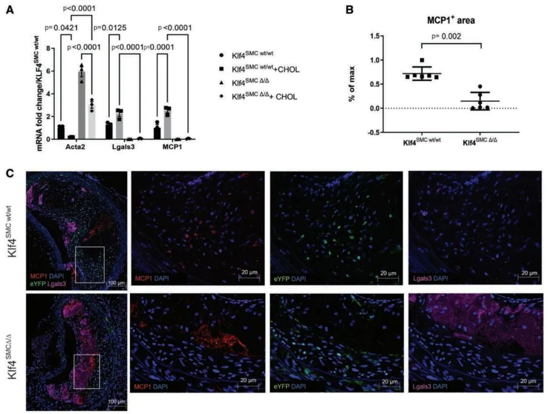
First, to investigate whether SMCs express MCP1 under atherosclerotic pathological conditions, the authors conducted RNAscope fluorescence in situ hybridization analysis on BCA (brachiocephalic artery) sections from wild-type and ApoE-/- mice. The results showed significant fluorescence staining in the vascular media and endothelial layers of ApoE-/- mice, and cultured mouse aortic SMCs significantly upregulated MCP1 transcription after 3 days of cholesterol treatment. Additionally, SMC-specific knockout of Klf4 reduced MCP1 production. Moreover, MCP1 protein expression was significantly reduced in the BCA lesion plaques of Klf4 knockout mice. Therefore, MCP1 is expressed in SMCs in a Klf4-dependent manner.
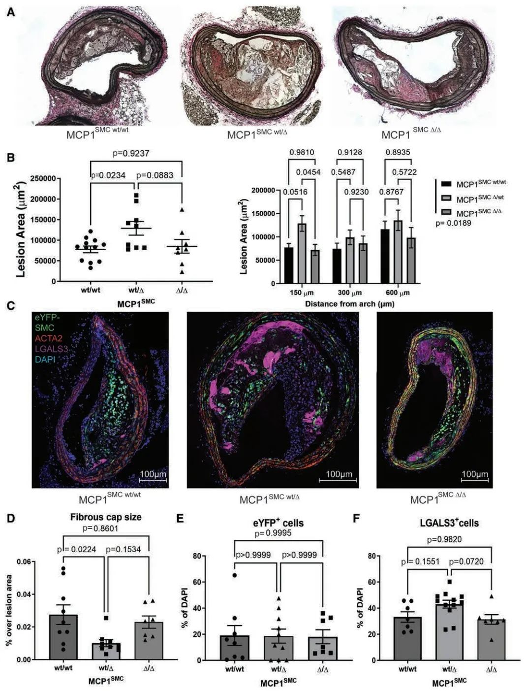
To explore whether SMCs influence the progression of atherosclerosis by secreting MCP1, the authors used lineage tracing technology to cross Myh11-CreERT2 Rosa-eYFP ApoE-/- mice with MCP1-mCherryflox/flox mice. The resulting positive mice were injected with tamoxifen at 6 weeks of age to induce MCP1 knockout in SMCs and were continued to be fed for 18 weeks to induce atherosclerotic plaque formation. Surprisingly, compared to wild-type controls and homozygous MCP1SMC Δ/Δ mice, heterozygous MCP1SMC wt/Δ mice showed increased BCA lesion area, decreased fibrous cap thickness, and an increased number of LGALS3+ macrophages in the lesions. The authors speculate that the increased expression of MCP1 produced by non-SMCs in homozygous MCP1SMC Δ/Δ mice may account for this. Therefore, the heterozygous deletion of MCP1 in SMCs exacerbated the progression of atherosclerosis.
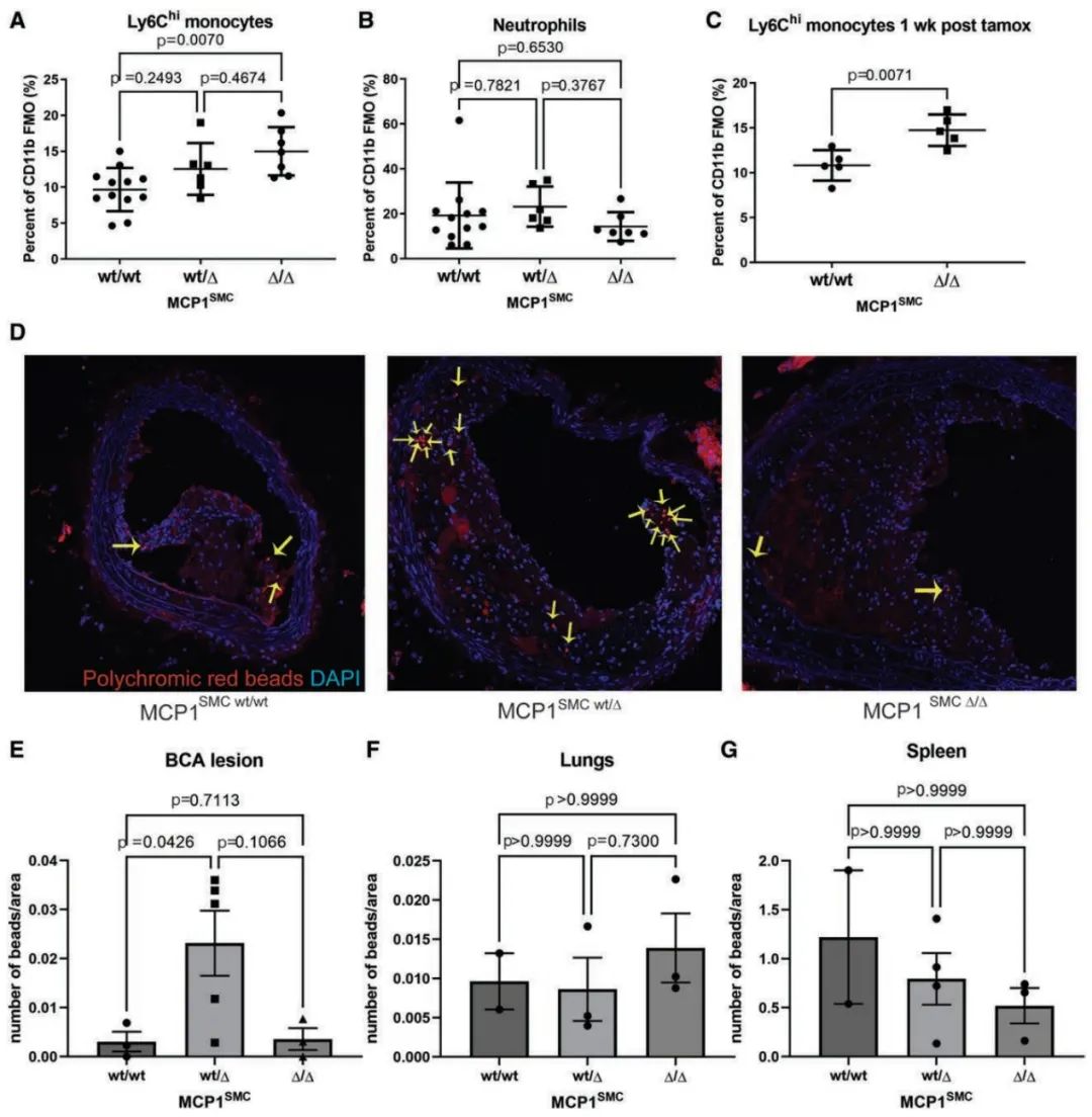
MCP1 is known to influence the migration of Ly6Chi inflammatory monocytes. The authors collected peripheral blood samples from MCP1SMC mice after 18 weeks of feeding and examined the leukocyte content using flow cytometry. The results indicated that the absence of MCP1 in SMCs led to a dose-dependent increase of Ly6Chi monocytes in the peripheral blood. To further investigate the dynamics of monocyte recruitment and egress in MCP1SMC mice, the authors injected fluorescent beads into the tail vein of MCP1SMC mice at 16 weeks and measured the bead content in the lesions at 18 weeks. The results showed that the bead content in lesions of MCP1SMC wt/Δ mice was significantly increased, indicating increased recruitment of monocytes. Therefore, the increase in monocyte numbers in the blood after the absence of MCP1 is not due to a decrease in tissue recruitment. The specific knockout of MCP1 in SMCs is associated with increased levels of Ly6Chi monocytes in the peripheral blood, which may lead to aggravated atherosclerosis and increased macrophages in the lesions.
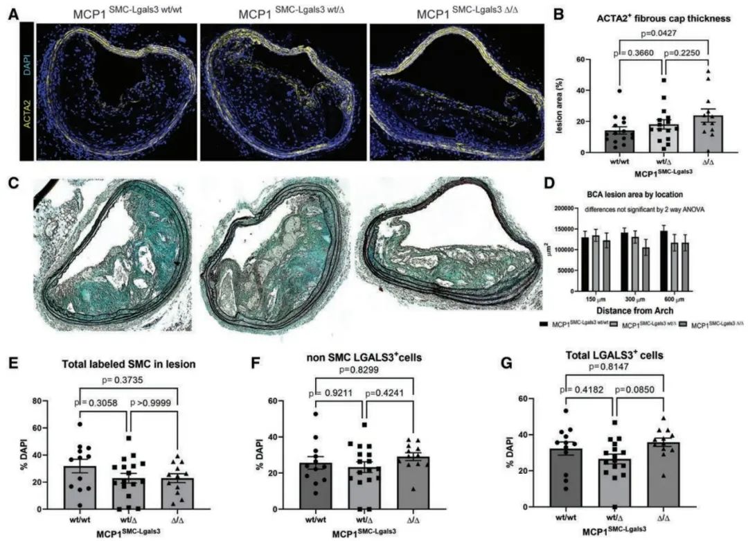
Subsequently, to explore the role of MCP1 produced by Lgals3+ SMCs in late-stage lesions, the authors crossed Myh11-DreERT2 Lgals3-Cre Rosa-tdTomato eGFP mice with MCP1flox/flox ApoE-/- mice and analyzed lesion size and plaque stability indicators after 18 weeks of feeding. The results showed that in MCP1SMC-Lgals3 Δ/Δ mice, the fibrous cap area of the plaque was significantly increased, while Ly6Chi monocytes in the aorta were significantly reduced. However, there was no overall change in lesion size among MCP1SMC-Lgals3 wt/wt, MCP1SMC-Lgals3 wt/Δ, and MCP1SMC-Lgals3 Δ/Δ mice. Furthermore, the knockout of MCP1 in Lgals3+ SMCs did not affect the total number of SMCs or the number of Lgals3+ cells. Therefore, the knockout of MCP1 in Lgals3+ SMCs had an opposite phenotype compared to the knockout of MCP1 in Myh11+ SMCs.
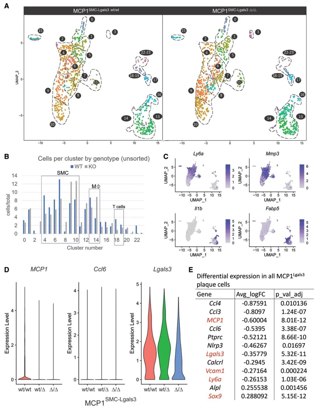
Finally, to investigate the effects of MCP1SMC-Lgals3 knockout on various phenotypes of SMCs and non-SMCs in late-stage atherosclerosis, the authors performed scRNA-seq on late BCA lesion plaques. The results indicate that MCP1 produced by Lgals3+ SMCs influences late-stage atherosclerotic plaques by regulating SMC phenotypes as well as multiple macrophage subpopulations and T cells.
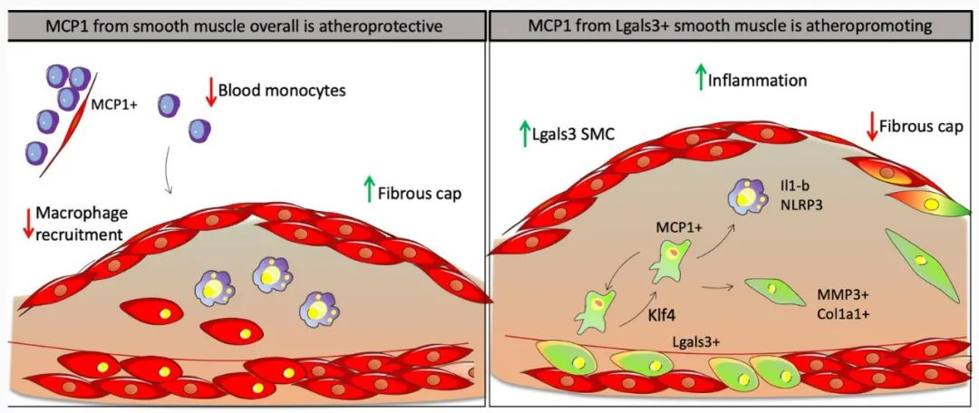
In conclusion, the authors demonstrated through SMC-specific MCP1 knockout that SMCs are the primary source of MCP1 in vivo, and the absence of MCP1 in all Myh11+ SMCs leads to an increase in circulating Ly6Chi monocytes, which is favorable for plaque formation. Furthermore, MCP1 produced by Lgals3+ SMCs has a detrimental effect on plaque lesions by prolonging chronic inflammation and promoting SMC phenotype transition, leading to reduced fibrous caps. This study uncovers a new mechanism of SMC phenotype transition and provides new potential intervention targets for the prevention and treatment of atherosclerosis.
https://www.ahajournals.org/doi/full/10.1161/ATVBAHA.122.317882?rfr_dat=cr_pub++0pubmed&url_ver=Z39.88-2003&rfr_id=ori%3Arid%3Acrossref.org
[Cardiovascular Insights]: This public account reports the latest research progress in the cardiovascular field both domestically and internationally. We welcome you to follow, share, and submit contributions. For submissions, please contact:13853112673 (same as WeChat, please choose this first,thank you very much) or [email protected]. If you like this article, please “like+ view” to support, let cardiovascular knowledge spread more!!