
Impact Factor: 7.7
Research Overview: Pulmonary hypertension (PH) is a heterogeneous group of diseases defined as a mean pulmonary artery pressure ≥20 mmHg measured during right heart catheterization at rest. Pulmonary arterial hypertension (PAH) is one of the main subtypes of PH, characterized by pulmonary vascular remodeling mediated by excessive proliferation of pulmonary artery smooth muscle cells (PASMCs). Although vasodilators are currently the main treatment for PAH, they have been shown to be unable to reverse smooth muscle cell proliferation and are expensive. Therefore, discovering new pathological mechanisms of PAH is crucial for developing therapeutic targets against potential vascular remodeling. For this purpose, the authors integrated four PAH datasets and conducted a comprehensive bioinformatics analysis.

The authors first identified differentially expressed genes (DEGs) in PAH; then, they performed enrichment analysis on the DEGs from each dataset. Immune cell infiltration analysis revealed the association between the core genes of PAH and the immune system. The authors then further utilized machine learning to construct a PAH diagnostic model based on core genes and found that MACC1 is a potential therapeutic target for PAH. MACC1 was originally discovered as a gene associated with tumor metastasis (MACC1 can induce tumor occurrence, progression, and metastasis), but in this article, the authors focus on studying the relationship between MACC1 and PAH through data analysis and in vitro and in vivo experiments.
Please scan the QR code below

Research Results:
Identification of PAH Differentially Expressed Genes
The authors first collected the raw data from two GEO datasets, GSE113439 and GSE117261, to find PAH differentially expressed genes, identifying a total of 201 genes, of which 157 were upregulated and 44 were downregulated (Figure 1A-D; Figure C shows the intersection of upregulated DEGs; Figure D shows the intersection of downregulated DEGs).
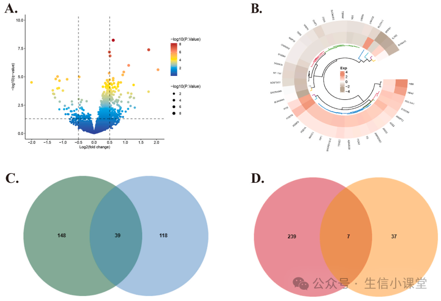
Next, the authors used the RRA (robust rank aggregation) algorithm (which considers not only the occurrence of genes but also their ranking in the list) and found that POSTN, ANGPT2, and PI15 were the top three upregulated DEGs. Among the downregulated genes, MT-TW ranked first, followed by MS4A15 and SOSTDC1.
Subsequently, the authors performed GSEA analysis on the DEGs’ enrichment pathways, revealing that DEGs were significantly enriched in pathways related to glutathione metabolism, glycolysis, gluconeogenesis, and T cell receptor signaling, while neuroactive ligand-receptor interaction Alzheimer’s disease-related pathways were suppressed.
Immune Infiltration Analysis
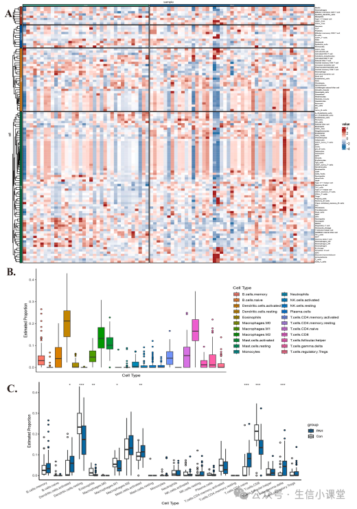
The authors further studied the differences in immune cell infiltration between PAH patients and healthy individuals using the IOBR and CIBERSORT algorithms. The results showed that in PAH patients, the infiltration levels of activated dendritic cells, mast cells, naïve CD4+ T cells, and follicular helper T cells were significantly higher than in healthy individuals. Resting dendritic cells, M1 macrophages, and CD8+ T cells were also found to play key roles in the progression of PAH (Figures 2A-C).
Key Gene Screening for PAH
Since the DEGs of PAH are enriched in multiple pathways and PAH has been confirmed to be related to immune infiltration, the authors further sought to find the core genes playing key roles using the WGCNA method. Through WGCNA analysis, the authors established 17 different co-expression modules; by studying the module-trait relationships, several modules were found to be associated with PAH (Figures 3A-C): the brown, blue, and turquoise modules showed close associations with certain immune cells. The authors then took the intersection of these three modules with DEGs, resulting in 17 intersection genes (Figure 3D).
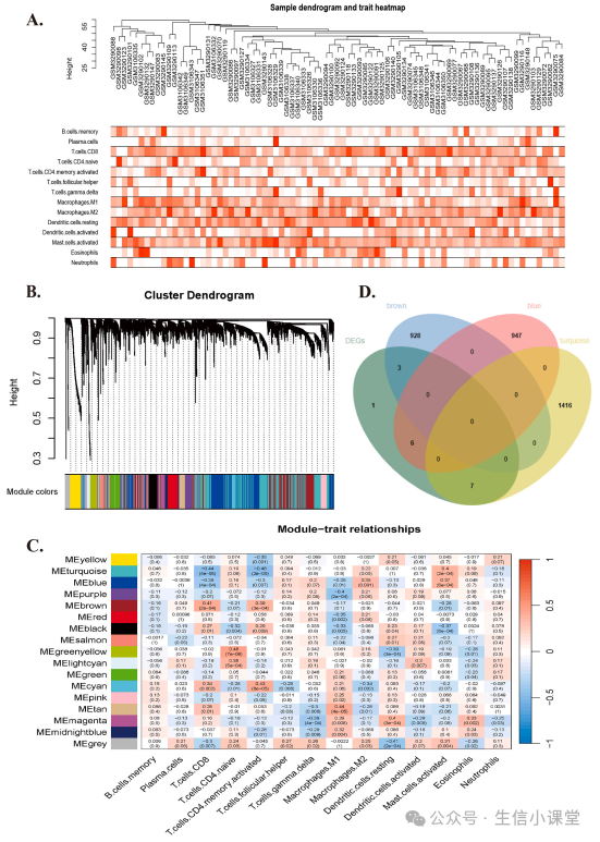
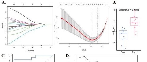
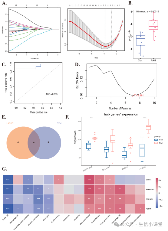
Furthermore, the authors used LASSO regression and SVM-RFE algorithms to narrow down the 17 intersection genes to 4 (the intersection of LASSO and SVM), which were identified as diagnostic biomarkers for PAH (Figures 4A-E). To verify the expression of these 4 genes in PAH, the authors used a third PAH dataset GSE53408. The results showed that these 4 genes were all upregulated in PAH compared to normal samples (Figure 4F). Additionally, these 4 genes were associated with certain immune cells, such as CD8+ T cells, CD4+ T cells, M2 macrophages, etc. (Figure 4G).
Clinical Data Validation
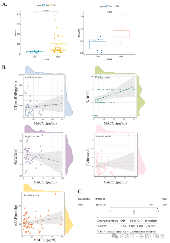
The authors first used the last public dataset GSE22356 to validate the upregulation of MACC1 in the serum of PAH patients (Figure 5A); then collected blood samples from 59 PAH patients and 22 healthy controls to determine serum MACC1 levels. ELISA results showed that serum MACC1 levels in PAH patients were significantly higher than in the healthy control group (p=0.012; Figure 5A). The authors also further analyzed the relationship between serum MACC1 levels and clinical characteristics of PAH patients, including WHO functional classification, N-terminal pro-B-type natriuretic peptide (NT-proBNP) concentration, pulmonary vascular resistance (PVR), mean pulmonary artery pressure (mPAP), and 6-minute walk distance (6MWD). The results showed that serum MACC1 levels were positively correlated with WHO functional classification, NT-proBNP concentration, PVR, and mPAP; while negatively correlated with 6MWD (Figure 5B). Logistic regression analysis still showed that MACC1 concentration is a risk factor affecting clinical PAH (Figure 5C).
In Vivo and In Vitro Experimental Validation
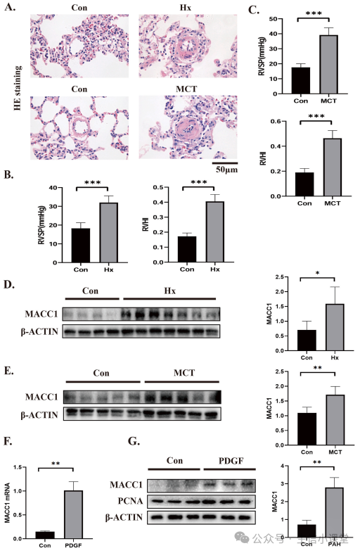
Finally, the authors conducted experiments to validate the role of MACC1 in PAH. First, the authors constructed PAH rat models using two methods. PAH: 10 rats were randomly divided into 2 groups: control group (n = 5) and MCT group (n = 5). The MCT group was induced with pulmonary arterial hypertension by intraperitoneal injection of 60 mg/kg body weight of monocrotaline (MCT). Hypoxia: 10 rats were randomly divided into control group (n = 4) and hypoxia group (n = 6), where the hypoxia group was given 10% oxygen, and pulmonary arterial hypertension developed after 3 weeks. Both methods effectively constructed the PAH animal models (Figures 6A-C; right ventricular systolic pressure (RVSP) and right ventricular to left ventricular plus septal weight ratio (RV/LV + S) were significantly increased in rats treated with MCT and hypoxia). The collected lung tissue homogenates also showed significantly high expression of MACC1 in the PAH group (Figures 6D-E). As mentioned earlier, excessive proliferation of PASMCs is the primary cause of pulmonary vascular remodeling. Therefore, the authors isolated rat PASMCs and stimulated them with platelet-derived growth factor BB (PDGF-BB) (PDGF-BB is a cytokine considered a key mediator of PASMC proliferation in PAH progression). After PDGF-BB stimulation, activation of MACC1 expression was observed (Figures 6F and G)—the mRNA/protein expression levels of MACC1 and proliferating cell nuclear antigen (PCNA—a proliferation marker) protein levels were significantly increased.
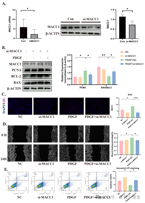
Knockdown of MACC1 Enhances PASMCs Proliferation, Migration, and Resistance to Apoptosis
To further elucidate the role of MACC1 in PASMCs, the authors knocked down MACC1 at the transcriptional level to assess whether reducing MACC1 levels would improve the malignant phenotype of PASMCs (proliferation, migration, and resistance to apoptosis) (Figure 7A). The results showed that MACC1 knockdown led to downregulation of PDGF-BB-induced PCNA expression and EDU levels, while upregulating the expression of apoptosis markers BAX/Bcl-2 (Figures 7B and C). These findings suggest that MACC1 plays a key role in PDGF-BB-induced PASMC proliferation and apoptosis resistance. The authors then used scratch assays to confirm that migration of PASMCs significantly decreased after MACC1 knockdown (Figure 7D). Finally, flow cytometry observed reduced apoptosis of PDGF-BB-induced PASMCs, while siMACC1 + PDGF group showed increased apoptosis (Figure 7E). In summary, these results indicate that MACC1 may promote the transition of PASMCs to a malignant phenotype (proliferation, migration, and resistance to apoptosis) under PDGF-BB induction.
Research Summary:
The authors first analyzed and screened DEGs from two PAH datasets using RRA and Limma methods. Based on WGCNA and machine learning methods, MACC1 was selected for further study in independent datasets. MACC1 was not only elevated in in vivo and in vitro experiments but also increased in the serum of clinical PAH patients. In vitro experiments with MACC1 knockout PASMCs also showed a reduction in malignant phenotype (proliferation, migration, and resistance to apoptosis). The greatest innovation of this article lies in the exploration of MACC1’s role in PAH. Previous studies have shown that MACC1 is overexpressed in most solid cancers and is directly related to tumor metastasis and prognosis. Although MACC1 plays a role in cancer progression, and the related HGF/c-Met receptor pathway is considered closely related to PASMC proliferation, no research has been conducted on its role in PAH. Through a series of bioinformatics and experimental analyses, the authors revealed that MACC1 could become a promising biomarker for early diagnosis and treatment of PAH.
————Divider————
Love me and ⭐star⭐ me. I originally thought that the so-called “WeChat revision,” “not starring will not receive push notifications,” etc., were just excuses for public accounts to seek everyone’s care! I didn’t expect it! I didn’t expect! Heartbreaking! This is actually true! You really won’t receive push notifications! So! If dear little friends feel that “Bioinformatics Classroom” helps you a little bit, I sincerely ask everyone: ⭐star⭐ hee hee hee! Thank you!