
Diffusion refers to a microscopic, random motion of molecules caused by thermal energy, also known as Brownian motion.
The different structures of human tissues lead to variations in the restrictions on the diffusion movement of water molecules in different directions. If the restricted diffusion of water molecules is symmetrical in all directions, it is called isotropic diffusion; if it is asymmetrical, it is called anisotropic diffusion.
Anisotropic diffusion is commonly found in human tissues, particularly in the white matter nerve fiber bundles, where water molecules diffuse more freely along the long axis of the nerve fibers, while diffusion in the direction perpendicular to the long axis is restricted by cell membranes and myelin.
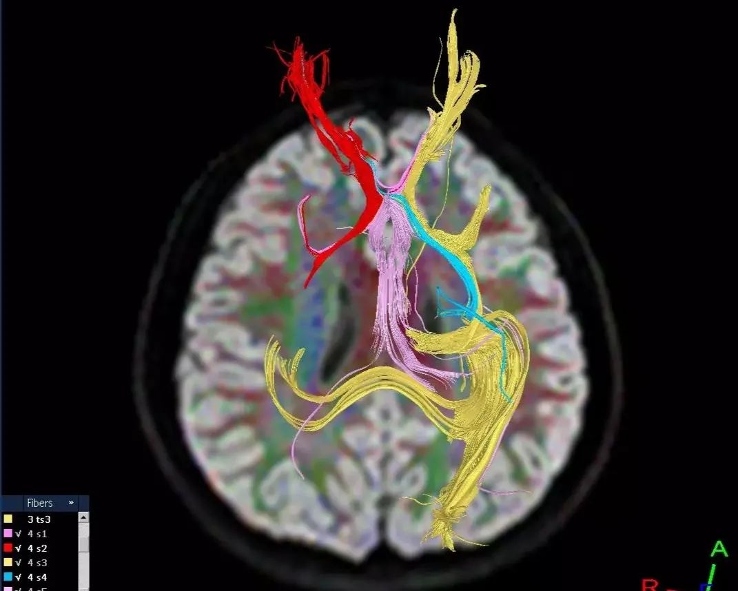
If a diffusion-sensitive gradient field is applied in more than 6 directions, the anisotropy of water molecule diffusion in each voxel can be detected. This technique is known as diffusion tensor imaging (DTI), which plays a significant role in reflecting the orientation of white matter fiber bundles in neuroscience research.
Through DTI analysis, the molecular mean diffusivity (MD) or apparent diffusion coefficient (ADC), fractional anisotropy (FA), axial diffusivity (the diffusion rate along the principal diffusion direction, AD), and radial diffusivity (RD) can be inferred for each voxel.
Related Concepts
1. Diffusion Coefficient (diffusion coefficient, DC): Indicates the range of free diffusion of molecules per unit time.
2. Diffusion Sensitivity Factor b-value (b value): Reflects the sensitivity of MRI imaging sequences (such as SE, FSE, EPI) to diffusion motion, representing the ability of the imaging sequence to detect diffusion.
3. Apparent Diffusion Coefficient (apparent diffusion coefficient, ADC): Describes the speed and range of diffusion motion of water molecules in different aspects during magnetic resonance diffusion-weighted imaging.
Clinical
1. Diffuse Axonal Injury (diffuse axonal injury, DAI)
2. Leukoaraiosis (leukoaraiosis)
3. Wallerian Degeneration (wallerian degeneration, WD)
Wallerian degeneration is the anterograde degeneration of axons, where the myelin sheath of the nerve axon disintegrates following damage to adjacent axons or the death of nerve cells. It is most commonly secondary to ipsilateral cerebral infarction, where the corticospinal tract exhibits WD. DTI is more sensitive than T2-weighted imaging in detecting WD.
4. Development, Maturation, and Aging of the Brain (developing brain, maturation, and aging)
5. Cerebral Ischemia (cerebral ischemia)
When cerebral blood flow drops below 10-15 ml/100 g/min, it leads to an increase in intracellular water volume, causing water to flow from the interstitial space into the cells, resulting in cytotoxic edema. Traditional MR imaging often struggles to detect acute cerebral infarction; the extent of ischemic brain tissue is only identifiable at a later stage when vasogenic edema appears. DWI and DTI can detect acute cerebral infarction earlier than conventional MR imaging when it appears normal.
