
Magnetic Resonance Diffusion Weighted Imaging (DWI) is the only non-invasive imaging technique that can detect the diffusion of water molecules in vivo, and it is also the most widely used magnetic resonance functional monitoring technique in clinical applications.
The diffusion sequence (technique) is not only widely used in clinical diagnosis with magnetic resonance but is also quite common in research papers; every year, diffusion-related papers are the most numerous at various imaging conferences.


Figure 1-2: The number of diffusion-related papers is the highest every year.
Given the importance of the diffusion sequence in both clinical and research contexts, it is essential to perform diffusion imaging effectively. So, how can we achieve this?
To perform diffusion imaging well, we first need to understand what a diffusion sequence is, its characteristics, and which parameters determine the contrast and quality of diffusion images.
1. Overview of Diffusion
Diffusion, in English, can be translated as: diffusion, spreading. The process of diffusion or spreading movement refers to the random, irregular motion of molecules (mainly water molecules in magnetic resonance) driven by temperature, which is known as Brownian motion.
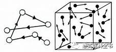
Figure 3: Schematic diagram of the diffusion movement of water molecules.
What rules does the diffusion movement of water molecules in magnetic resonance follow? Einstein proposed an equation: <X>2=2DTd. In this equation, the distance (magnitude) of molecular diffusion or diffusion displacement is positively correlated with the diffusion time (measurement time) and the tissue diffusion coefficient. D is approximately 10-3mm2/s, Td is 50ms, x=10μm, meaning that the diffusion distance of free water is about 10μm, which is on the same order of magnitude as many structures in the human body. Of course, diffusion is also affected by temperature. However, in in vivo magnetic resonance examinations, this factor can be relatively disregarded.
The diffusion technique can detect the diffusion of water molecules in vivo to assess tissue conditions and infer microscopic morphological and structural characteristics.
After understanding the basic principles of diffusion, we should know how the diffusion sequence is constructed and how it differs from other traditional weighted sequences.
2. Principles of DWI Sequences
Compared to conventional weighted sequences, the diffusion sequence detects the diffusion of water molecules by applying a diffusion gradient field on the basis of traditional sequences.
This diffusion gradient is also known as a bi-polar gradient. What does this mean? It means that the correlation of this gradient is opposite; through this bi-polar gradient, the diffusion of water molecules can be detected.
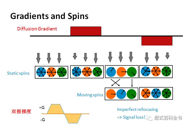
Figure 4: Principles of DWI sequence and schematic diagram of bi-polar gradient.
Under normal circumstances, stationary tissues (protons) experience the effects of two leaves of the bi-polar gradient, and the phase loss between protons cancels out precisely, collecting signals at TE time, achieving the same phase, maximizing the signal without decline.
However, for moving tissues (protons), because they are also in motion during the time of the two bi-polar gradients, the phase loss of protons cannot be fully compensated, leading to reduced signals. The faster the motion, the less the phase loss of protons can be compensated, and the more significant the signal reduction.
By using the bi-polar gradient, it is easy to detect stationary and moving tissues.
The bi-polar gradient can be combined with spin echo sequences or gradient echo sequences.
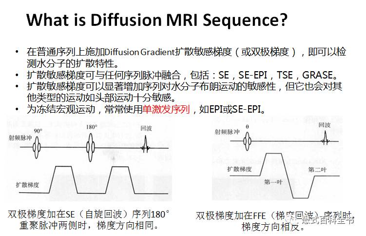
Figure 5: DWI sequence situation.
If combined with the SE spin echo sequence, the two leaves of the bi-polar gradient are generally placed on either side of the 180° refocusing pulse. In this case, the directions of the two bi-polar gradients are the same because the 180° refocusing pulse reverses the phase position of the protons, and then the phase loss is compensated by the same phase bi-polar gradient.
Conversely, if combined with the FFE gradient echo sequence, since there is no 180° refocusing pulse, the directions of the bi-polar gradients are opposite; thus, one positive and one negative can compensate for the phase difference of stationary protons.
Another characteristic is that the diffusion sequence is generally read out using EPI, which allows for faster scanning times.
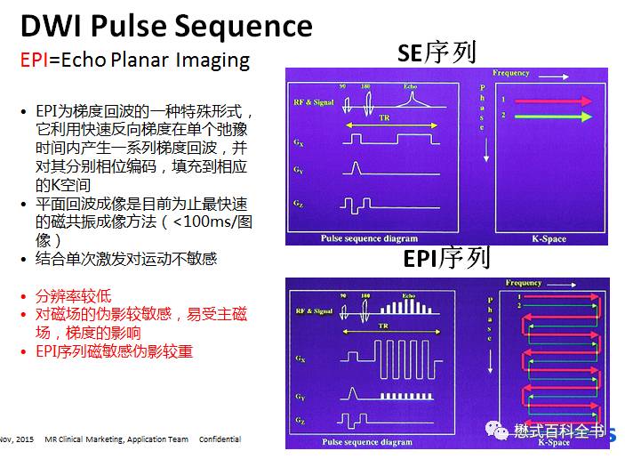
Figure 6: EPI readout method.
The use of EPI for DWI has the significant advantage of speed and is insensitive to gross motion artifacts. Of course, EPI readout can also produce many unique artifacts and defects.
First, EPI quickly reads out signals by switching positive and negative gradients, which can easily accumulate phase differences, leading to image distortion; moreover, if the echo train length increases, the EPI factor can also exacerbate EPI artifacts and distortion. Additionally, the EPI sequence does not use multiple 180° refocusing pulses to gather signals, making it sensitive to magnetic susceptibility and prone to magnetic susceptibility artifacts.
3. Characteristics and Parameters of DWI Sequences
The commonly used DWI sequence in clinical practice is SE-EPI, which consists of a spin echo followed by fast signal readout using EPI, primarily based on single-shot excitation.
What is single-shot excitation? It means that a single RF pulse is used to excite and complete an image acquisition; theoretically, the TR time is infinite.
Since it is primarily based on single-shot excitation, it cannot demand excessive resolution (large matrix) because it cannot complete all phase-encoded lines within a single TR (if the resolution is too high).
This leads to the following characteristics of clinical DWI sequences and images:
1. Limited resolution;
2. Image distortion (occurring in the phase encoding direction);
3. Chemical shift also occurs in the phase encoding direction (in conventional sequences, chemical shift occurs in the frequency encoding direction);
4. Sensitive to magnetic susceptibility, prone to magnetic susceptibility artifacts;
5. During the sequence scanning process, gradient oscillation is frequent, resulting in high noise levels.
To address these characteristics, different manufacturers have devised various methods to improve the diffusion sequence, which I will discuss later.
In addition to these, the most crucial parameter of the diffusion sequence that everyone has heard of is called diffusion sensitivity, also known as the B-value.
Here, let me ask everyone why this value is called the B-value and not the C-value or D-value? It is said to commemorate the French founder of the diffusion sequence, Denis Le Bihan, hence the name B-value.

Figure 7: The founder of the diffusion sequence, Denis Le Bihan.
Most teachers using the DWI sequence know that the higher the B-value, the greater the sensitivity of the diffusion sequence for detection, but the signal-to-noise ratio (SNR) decreases. But why is that? Many people may not fully understand.
First, let’s look at the definition of the B-value.
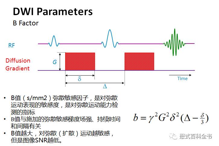
Figure 8: The significance of the B-value.
The formula for the B-value, as shown in the figure above. The B-value reflects the magnitude (effectiveness) of the applied diffusion gradient. In the formula, G represents the amplitude of the diffusion gradient, δ represents the duration of a single gradient application, and Δ represents the time interval between two diffusion gradients.
Therefore, we know that a larger B-value reflects a stronger diffusion gradient. The B-value is positively correlated with G, δ and Δ.
So, how can we increase the B-value using the above methods?
First, we can increase G, that is, increase the amplitude of the diffusion gradient field; of course, this method cannot increase the B-value indefinitely, as physical factors limit the diffusion gradient field from being increased indefinitely. When the gradient field reaches a maximum value, it cannot be increased further.
Secondly, we can increase the B-value by increasing δ. This method is feasible. When the diffusion gradient field reaches its maximum value, increasing the duration can enhance the effectiveness of the diffusion gradient.
Thirdly, we can achieve this by increasing Δ.
Now, at this point, it is not difficult to understand why the larger the B-value, the lower the SNR of the image. If you still do not understand, take a look at the schematic diagram.
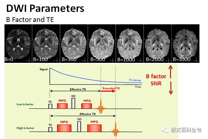
Figure 9: The relationship between B-value size, TE, and SNR.
Due to the size of the diffusion gradient field being limited (maximum value), when the B-value continuously increases, it is generally achieved by increasing the duration of the diffusion gradient or the time interval between the two gradients.
Thus, to increase the duration of the diffusion gradient or the time interval between the two gradients, the TE acquisition time must be extended.
When other factors remain unchanged, extending TE will naturally decrease the SNR, which also explains the physical reason why the larger the B-value, the lower the SNR.
Another important parameter is called the EPI factor. This can be translated as the EPI factor. What does it mean? Since most DWI sequences used in clinical practice are single-shot, meaning that the entire K-space filling must be completed in one acquisition; theoretically, the TR value remains unchanged. The time to complete an image does not change.
If we increase the resolution, the number of acquisition points and phase encoding steps will increase, meaning that the number of echoes to be acquired in one shot will also increase, which will inevitably increase the EPI factor.
As mentioned earlier, if the EPI factor is too large, it will exacerbate phase accumulation errors, causing image distortion.
Therefore, to improve image quality in the DWI sequence, one crucial factor is to reduce the EPI factor as much as possible.
So, how can we reduce the EPI factor?
1. We can reduce the resolution (matrix); this way, fewer phase encoding steps will lead to a lower EPI factor (this is also why the DWI sequence does not have high resolution);
2. Use parallel acquisition techniques, such as SENSE technology, to reduce the number of phase encoding lines acquired;
3. Use half-scan techniques to reduce the number of phase encoding lines acquired.
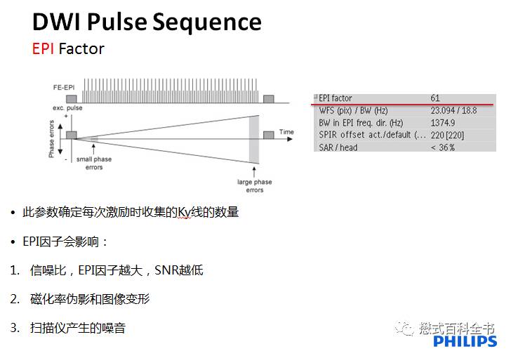
Figure 10: How EPI factor affects image quality.
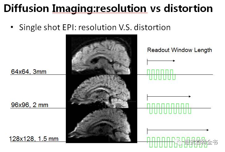
Figure 11: Lower resolution leads to smaller EPI factor and less image distortion.
To perform DWI sequences effectively, we must consider reducing the EPI factor and preventing artifacts related to DWI sequences.
Additionally, DWI sequences generally require fat suppression. This is because fat and water have chemical shifts, and diffusion sequences are prone to distortion. If fat tissue is not suppressed, the chemical shift artifacts and image distortion of the DWI sequence will be more severe, affecting image diagnosis.
4. Artifacts Related to DWI Sequences
Due to the characteristics and principles of the DWI sequence, it is prone to many artifacts. First, there is image distortion, as mentioned above. Additionally, there are some artifacts unique to diffusion sequences.
N/2 ghost artifacts: Such artifacts are rarely seen on new machines. They arise from insufficient sampling; in DWI-EPI sequences, the rapid switching of bi-polar gradients during signal acquisition leads to imprecise phase information.
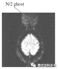
Figure 12: N/2 ghost artifact.
Magnetic susceptibility artifacts: Due to the use of EPI readout in DWI sequences, with few (or no) 180° refocusing pulses, they are highly sensitive to magnetic susceptibility, creating significant magnetic susceptibility artifacts at tissue boundaries and regions of magnetic susceptibility variation.
Chemical shift artifacts: In conventional sequences, non-fat-suppressed sequences may exhibit chemical shift artifacts, but they are generally mild and occur in the frequency encoding direction. However, in diffusion sequences, due to low resolution and the use of EPI readout, chemical shift artifacts occur in the phase encoding direction, leading to voxel displacement and affecting image observation and accurate measurement of ADC values.
5. Clinical Applications and Considerations of DWI Sequences and ADC Values
Why can’t we only look at DWI images to judge diffusion restriction? We also need to consider ADC images.
6. Clinical Applications of DWI
Currently, DWI sequences are applied to various parts of the body, with the most common uses being in the brain, abdomen, and pelvis.
The application in the brain is relatively simple, using the traditional SE-EPI sequence, completing a 2D whole-brain scan in 15-40 seconds, and then generating an ADC image to determine whether there is diffusion restriction and signal abnormalities based on the DWI and ADC images.
In abdominal applications, the liver moves up and down and rolls forward and backward. Therefore, when performing abdominal DWI, we need to consider how to freeze respiratory motion.
How to freeze respiratory motion during magnetic resonance scanning?
For abdominal DWI sequences, depending on different manufacturers and hospital practices, there are several methods:
1. Use BH, a segmented breath-hold scanning method for abdominal diffusion. This method is more commonly used by GE users. The advantage is: fast scanning; the disadvantage is: unstable image quality, sometimes poor.
2. Use RT, a respiratory-triggered method for abdominal DWI. This method combines the DWI sequence with RT technology, acquiring signals during the end-expiration plateau phase. The advantage is: if the patient’s breathing is uniform, the image quality is good; less image distortion; higher resolution; the downside is: longer scanning time; if the patient’s breathing is uneven, the effect may not be good. Many Philips users generally recommend this.
3. Completely free-breathing scanning. In DWI sequences, due to the use of EPI, the acquisition time is very fast and is insensitive to gross motion artifacts. Sometimes, abdominal DWI can be scanned entirely with free breathing (without any respiratory compensation technology). The advantage is: scanning speed is faster than respiratory triggering; image quality is better than breath-hold scanning. The disadvantage is: resolution may not be very high; sometimes there may be significant artifacts. In this case, generally, Philips and Siemens 1.5T users use this method more often.
Additionally, why does free-breathing scanning of abdominal DWI not cause significant motion artifacts? Currently, it is believed that the overall respiratory motion mainly affects the incoherent motion within a voxel (IVIM) and does not influence the phase loss caused by hydrogen proton diffusion motion.
7. How to Choose B-values for DWI Sequences
Different parts of the body use different B-values.
For brain scans, the B-value we typically use is 1000. Of course, the higher the B-value, the more sensitive it is to diffusion motion, but SNR decreases. Therefore, in the brain, B-values higher than 1000 may improve the detection rate for less sensitive lesions, but compared to the decline in SNR, the increase in efficacy is not significant. Thus, the standard adopted by most hospitals in China is to use a B-value of 1000 for brain DWI.
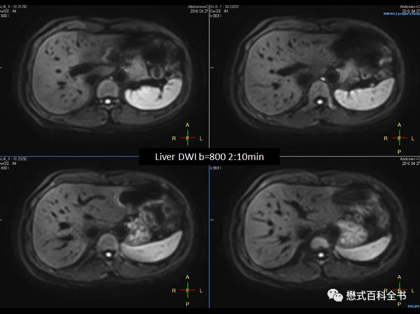
Figure 13: Abdominal DWI, B-value 800, scanning time 2 minutes 10 seconds, respiratory-triggered, clear image.
In the abdomen, currently, the consensus reached by most hospitals in China is:
In 1.5T, the abdomen uses two or three B-values, the highest B-value is 600, and a middle B-value can be set at 20 or 50. That is, three B-values: 0, 20 (50), 600;
In 3.0T, the abdomen routinely uses two or three B-values, the highest B-value is 800, and a middle B-value can be set at 20 or 50. That is, three B-values: 0, 20 (50), 800.
Someone may ask, what is the significance of a low B-value of 20 or 50?
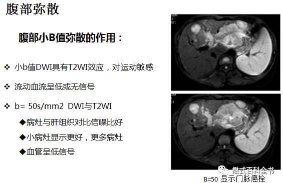
Figure 14: The significance of low B-values in diffusion.
Low B-values, due to their small size, have high SNR and are heavily T2-weighted. Moreover, due to the effect of the diffusion gradient (even with a low B-value), small blood vessels within the liver can be dispersed, allowing for the identification of small lesions and enhancing the contrast within the liver.
In the pelvis, high B-values for DWI are currently recommended clinically, especially for the prostate. I have performed many prostate diffusion scans and found that in 3.0T, if the B-value is less than 1500, the signal from the bladder often cannot be completely suppressed, which affects observation. Therefore, some hospitals use three B-values for prostate DWI: 0, 1000, 2000. Of course, as mentioned earlier, the larger the B-value, the lower the SNR. A B-value of 2000 may not provide sufficient SNR, necessitating an increase in NSA excitation times to improve the SNR. Additionally, considering the overall SNR, scanning time, and diffusion sensitivity, a high B-value between 1200-1500 can be considered.
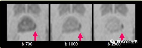
Figure 15: Prostate DWI with three B-values: 700, 1000, 2000. It can be observed that at a B-value of 2000, the signal of normal prostate tissue is suppressed, increasing sensitivity for detecting small lesions.
Additionally, some hospitals routinely perform spinal DWI scans in the sagittal position, recommending a B-value range of 500-800 to balance SNR and sensitivity.
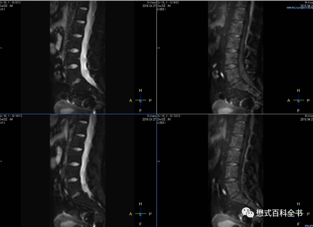
Figure 16: An example of lumbar DWI scanned from left to right, with B-values of 0, 800. Sagittal acquisition with minimal image distortion, performed on a 3.0T Philips Ingenia.
Furthermore, there is a special application for whole-body diffusion imaging. A certain company has termed this technology as a PET-like technique. However, I believe this naming is a bit inaccurate because the principles of whole-body diffusion DWIBS and PET are vastly different. This naming might confuse our own people and clinical doctors.
Whole-body background-suppressed diffusion DWIBS can be used for whole-body diffusion scanning, screening for tumors and the presence of systemic metastases.
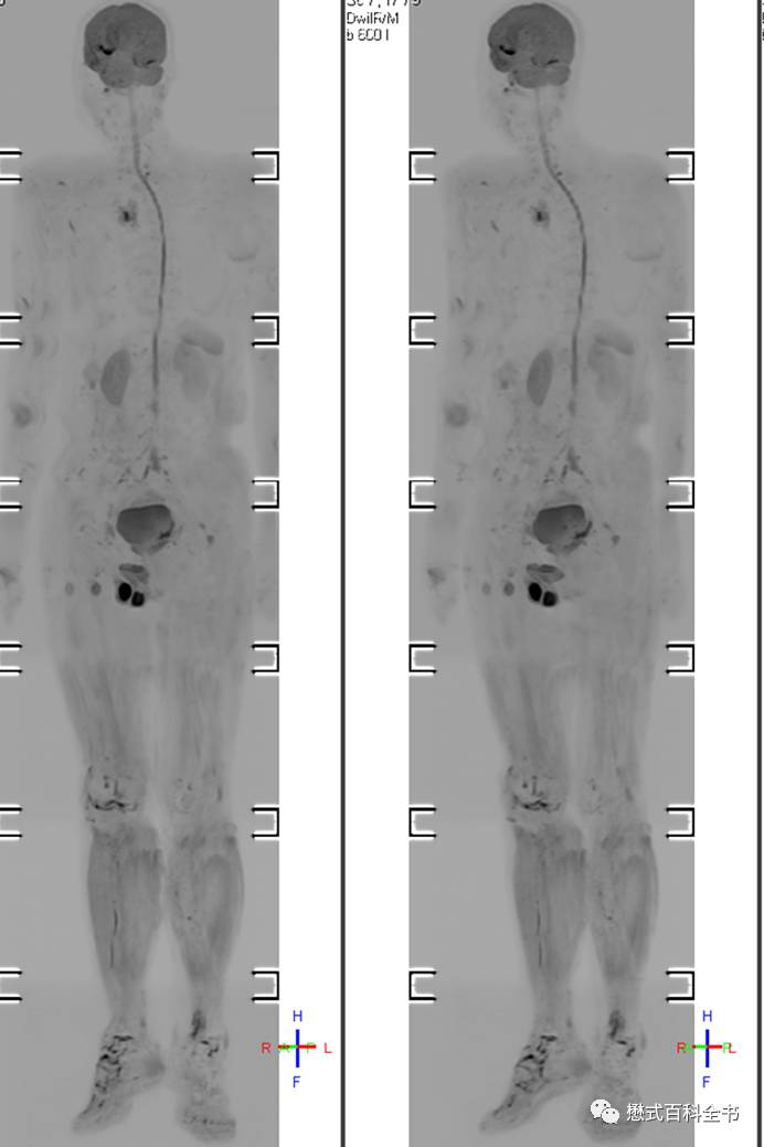
Figure 17: DWIBS whole-body background-suppressed diffusion.
8. Innovations and Improvements Based on the Characteristics of DWI Sequences
As mentioned earlier, conventional DWI sequences, primarily single-shot EPI acquisitions, lead to image distortion, low resolution, and sensitivity to magnetic susceptibility artifacts. So how can we improve the diffusion sequence through technology?
Philips has innovated the DWI-TSE sequence, which is a breakthrough improvement. We know that DWI sequences are generally acquired using EPI; however, Philips can use TSE (Turbo Spin Echo) sequences to create DWI sequences.
The TSE sequence, due to multiple 180° refocusing pulses, results in minimal image distortion and insensitivity to magnetic susceptibility artifacts.
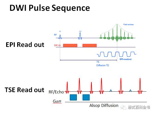
Figure 18: Traditional DWI-EPI diffusion sequence vs. Philips’ innovative DWI-TSE diffusion sequence.
Due to the use of TSE readout, image distortion is minimal, and it is insensitive to magnetic susceptibility artifacts.
The primary advantage is its application in magnetically sensitive areas, such as the cerebellopontine angle and neck regions.
Traditionally, due to the presence of a large amount of gas in these areas, magnetic susceptibility is exacerbated, leading to significant distortion and artifacts in conventional diffusion sequences, which are insufficient for clinical diagnosis; however, Philips’ DWI-TSE can effectively address these areas.
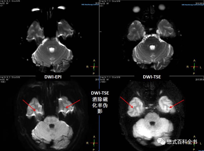
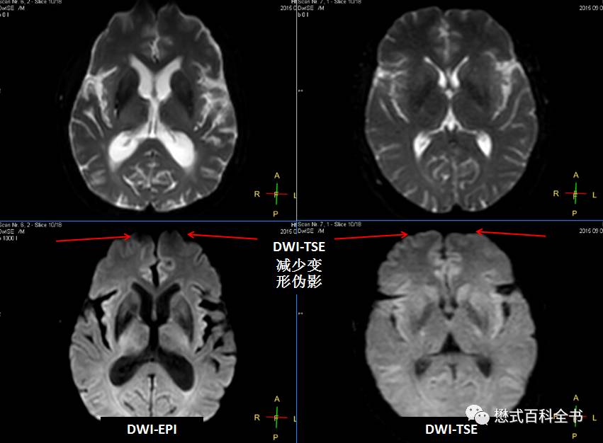
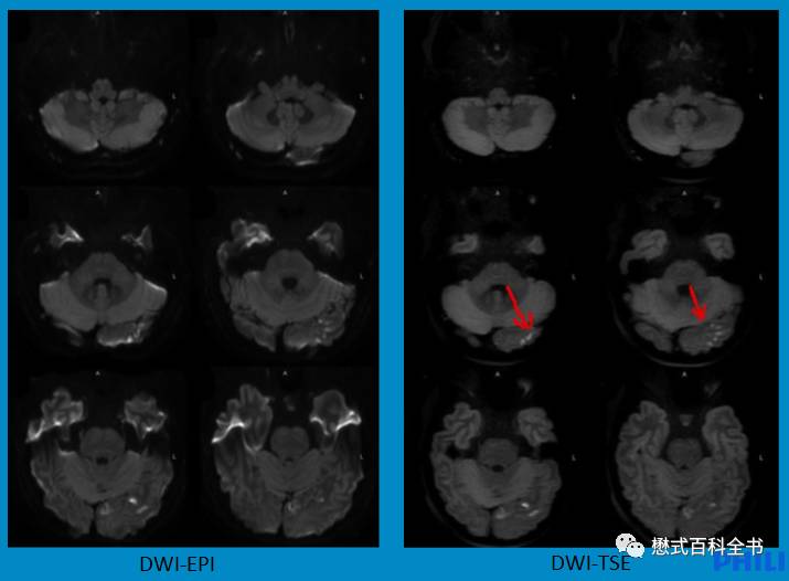
Figures 19-21: DWI-TSE sequence reduces diffusion image distortion and eliminates magnetic susceptibility artifacts, having very practical clinical significance for diffusion in the cerebellopontine angle and neck areas.
In addition to Philips, Siemens employs Resolve technology, which uses segmented readout in the readout direction to reduce the EPI factor.
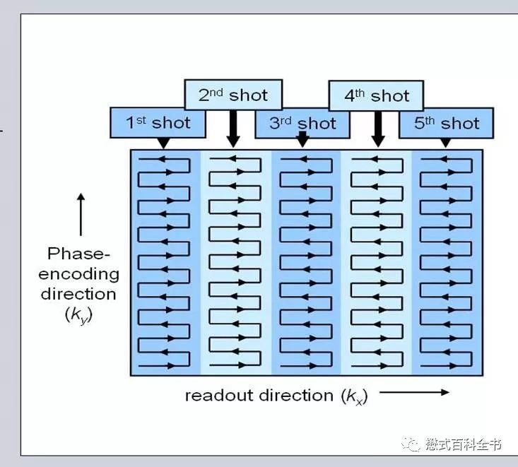
Figure 22: Schematic diagram and principle of Siemens Resolve DWI sequence.
This sequence uses serial segments in the readout direction, employing segmented K-space acquisition to reduce the EPI factor and minimize image distortion.
GE, in its DWI sequences, utilizes Propeller technology to eliminate gross motion artifacts and improve image quality.
Furthermore, to enhance the resolution of DWI sequences, all three companies (GPS) have focused on small field-of-view DWI. The small field-of-view is not the goal; the aim is to reduce voxel size and improve resolution.
Philips’ latest technology, Zoom DWI, ensures that when using a very small field of view for DWI sequences, there is no need for oversampling, preventing image distortion. As the field of view decreases without oversampling, if the matrix remains unchanged, this effectively reduces voxel size and enhances the resolution of DWI.
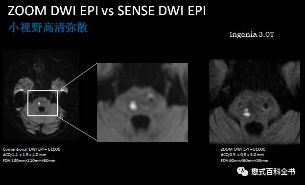
Figure 23: Zoom DWI, small field of view high-definition diffusion, improving resolution.
Siemens’ small field-of-view DWI technology is called ZooMit.
GE’s similar small field-of-view DWI technology is called FOCUS.
9. Innovations and Evolution of DWI Sequences
Traditional DWI sequences use single-shot DWI-EPI sequences, which have numerous shortcomings due to their design. To overcome these shortcomings, sequence designers have devised many solutions.
Single-shot sequences tend to have a large EPI factor, leading to image distortion. So, what if we use multi-shot? We could use DWI-EPI Multi-shot.
However, while multi-shot reduces image distortion, the scanning time increases significantly compared to single-shot, and the images become sensitive to motion artifacts, making this approach impractical.
Therefore, the focus shifted to reducing the EPI factor, which could only be achieved through acceleration, specifically by reducing the number of phase encoding steps.
For example: parallel acquisition techniques like SENSE, and half-scan techniques.
However, these techniques have a limit to their acceleration effects; beyond a certain point, artifacts will arise. Thus, to further enhance DWI image quality, this approach also has its limitations.
Consequently, Philips has approached the sequence design by addressing the EPI readout’s propensity for image distortion and phase accumulation errors by adopting TSE readout.
Siemens has focused on the readout method, addressing the large EPI factor by employing segmented K-space acquisition techniques in the readout direction.
Additionally, traditional DWI is based on a single-exponential model. Using the simplest two B-values, we can calculate the ADC value to measure the apparent diffusion coefficient.
In reality, this simplest model is overly idealistic.
It does not consider diffusion anisotropy or directionality. By adding these factors, we arrive at DTI;
It does not account for multi-exponential models. By incorporating this, we obtain the IVIM and stretched exponential models;
It does not consider the complexity of tissue structure; traditional diffusion models only consider Gaussian distribution cases and do not account for non-Gaussian distribution. By including this, we have DKI;
If we take into account the complexities of direction and structure, we also have DSI and NODDI.
Since continuing to write would delve into research areas beyond my clinical application training scope, I will refrain from writing further and will stop here.
I look forward to discussing diffusion models with everyone in the future.
The best way to support original content is: donations!