Written by | November
Edited by | Xi
Imaging of mRNA typically utilizes a method involving fluorescent proteins. For instance, green fluorescent protein is fused with the MS2 phage coat protein (MCP), forming MCP-GFP, which can be recruited to mRNA containing 24-48 consecutive MS2 hairpin structures in the 3′ UTR region【1】. This system is known as MS2-MCP. MCP can bind to the MS2 hairpin structure, and multiple repeated MS2 sequences can amplify the signal, allowing the observation of single mRNA signals under a fluorescence microscope. However, to remove unbound fluorescent proteins from the cytoplasm, a nuclear localization element is added to the MCP-GFP fusion protein, thereby reducing the fluorescent background in the cytoplasm and achieving better detection of mRNA signals. Nonetheless, this may introduce some false positive signals, as the labeled mRNAs contain dozens of nuclear localization sequences【2】.
To provide better mRNA monitoring tools, on August 31, 2019, the research group led by Samie R. Jaffrey at Cornell University (with Jiahui Wu as the first author) published an article titled Live imaging of mRNA using RNA-stabilized fluorogenic proteins in Nature Methods, introducing RNA-regulated conditional fluorescent proteins into the mRNA imaging process.
 RNA fluorescent aptamers are capable of binding to non-fluorescent molecules and converting them into fluorescent forms. When these fluorescent dyes are applied inside cells, RNA labeled by fluorescent aptamers can be detected through fluorescence microscopy【3】. However, the number of developed fluorescent aptamers is still limited, as very few aptamers are suitable for live cell imaging. Currently, most dyes are activated by lipids or DNA within the cells, leading to non-specific fluorescence【4】. To expand the application of fluorescent aptamer-based live cell imaging and address the issues brought by the MS2-MCP system, Jaffrey’s research group aims to find novel fluorescent dyes that can be genetically encoded. In most cases, fluorescent proteins are constitutively expressed and will be maintained in cells from the start; the authors aim to convert such constitutive fluorescent proteins into conditionally expressed proteins. Thus, the design focus is to find an mRNA monitoring system where excess proteins, other than those specifically bound by the RNA aptamer, will be degraded.
To find such RNA-regulated proteins, the first problem the authors need to solve is to identify a “destabilizing domain” that can regulate protein degradation, while this degradation can be suppressed by the binding of RNA aptamers. The authors designed a peptide segment with this dual function, named tDeg, which includes a degradation sequence and an arginine-rich RNA-binding peptide segment, Tat【5,6】 (Figure 1). tDeg allows the unstable characteristics of the protein to be eliminated in the presence of circular TAR RNA. When tDeg is expressed alone in HEK293T cells, almost no fluorescent signal can be detected. However, when circular TAR variant-2 RNA is introduced into the cells along with tDeg, the fluorescence intensity is 38 times higher than that of the control group RNA. Additionally, since the fluorescent RNA aptamer does not contain any cellular transport elements, the potential issues of the MS2-MCP system will no longer exist. Following the previous naming convention of RNA aptamers using colorful vegetables, the authors named the RNA aptamer in this study “Pepper”. The Pepper system can be used with different fluorescent proteins, making the system versatile with multiple color options. Furthermore, it can accurately indicate mRNAs with different subcellular localizations, further demonstrating the practicality of this system.
RNA fluorescent aptamers are capable of binding to non-fluorescent molecules and converting them into fluorescent forms. When these fluorescent dyes are applied inside cells, RNA labeled by fluorescent aptamers can be detected through fluorescence microscopy【3】. However, the number of developed fluorescent aptamers is still limited, as very few aptamers are suitable for live cell imaging. Currently, most dyes are activated by lipids or DNA within the cells, leading to non-specific fluorescence【4】. To expand the application of fluorescent aptamer-based live cell imaging and address the issues brought by the MS2-MCP system, Jaffrey’s research group aims to find novel fluorescent dyes that can be genetically encoded. In most cases, fluorescent proteins are constitutively expressed and will be maintained in cells from the start; the authors aim to convert such constitutive fluorescent proteins into conditionally expressed proteins. Thus, the design focus is to find an mRNA monitoring system where excess proteins, other than those specifically bound by the RNA aptamer, will be degraded.
To find such RNA-regulated proteins, the first problem the authors need to solve is to identify a “destabilizing domain” that can regulate protein degradation, while this degradation can be suppressed by the binding of RNA aptamers. The authors designed a peptide segment with this dual function, named tDeg, which includes a degradation sequence and an arginine-rich RNA-binding peptide segment, Tat【5,6】 (Figure 1). tDeg allows the unstable characteristics of the protein to be eliminated in the presence of circular TAR RNA. When tDeg is expressed alone in HEK293T cells, almost no fluorescent signal can be detected. However, when circular TAR variant-2 RNA is introduced into the cells along with tDeg, the fluorescence intensity is 38 times higher than that of the control group RNA. Additionally, since the fluorescent RNA aptamer does not contain any cellular transport elements, the potential issues of the MS2-MCP system will no longer exist. Following the previous naming convention of RNA aptamers using colorful vegetables, the authors named the RNA aptamer in this study “Pepper”. The Pepper system can be used with different fluorescent proteins, making the system versatile with multiple color options. Furthermore, it can accurately indicate mRNAs with different subcellular localizations, further demonstrating the practicality of this system.
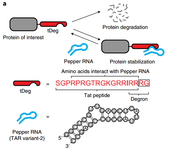
Figure 1: Schematic diagram of the Pepper-RNA regulated protein live imaging principle
In summary, the work of Jaffrey’s research group has found a system for real-time monitoring of mRNA that can more specifically use RNA aptamers to convert constitutive fluorescent proteins into conditionally luminescent fluorescent proteins. The RNA aptamer Pepper is used to control the stability of the fluorescent proteins; unbound fluorescents are rapidly degraded, while fluorescent proteins bound to Pepper RNA aptamers can stably exist. However, a key consideration for all systems using fluorescent proteins is the time required for fluorescent protein maturation (the kinetic properties of the fluorescent proteins themselves). Real-time monitoring of dynamic changes in mRNA in cultured cells or within animal cells will impose higher demands on the maturation time and other kinetic characteristics of fluorescent proteins in the mRNA live imaging system.
It is worth mentioning that on September 23, the research groups of Professor Yang Yi from the State Key Laboratory of Bioreactor Engineering and the Center for Interdisciplinary Research on Optogenetics and Synthetic Biology at East China University of Science and Technology, and Professor Zhu Linyong published an article titled Visualizing RNA dynamics in live cells with bright and stable fluorescent RNAs in Nature Biotechnology, which also developed the Pepper fluorescent protein (see BioArt report: NBT | Yang Yi/Zhu Linyong Team Develops Pepper Fluorescent Protein RNA).
Original link:
https://doi.org/10.1038/s41592-019-0531-7
1 Bertrand,E. et al. Localization of ASH1 mRNA particles in living yeast. Molecular Cell 2, 437-445 (1998).
2 Tyagi, S. Imaging intracellular RNA distribution and dynamics in living cells. Nature Methods 6, 331-338, doi:10.1038/nmeth.1321 (2009).
3 Paige, J. S., Wu, K. Y. & Jaffrey, S. R. RNA mimics of green fluorescent protein. Science 333, 642-646, doi:10.1126/science.1207339 (2011).
4 Fam, T. K., Klymchenko, A. S. & Collot, M. Recent Advances in Fluorescent Probes for Lipid Droplets. Materials 11, doi:10.3390/ma11091768 (2018).
5 Puglisi, J. D., Chen, L., Blanchard, S. & Frankel, A. D. Solution structure of a bovine immunodeficiency virus Tat-TAR peptide-RNA complex. Science 270, 1200-1203, doi:10.1126/science.270.5239.1200 (1995).
6 Ye, X., Kumar, R. A. & Patel, D. J. Molecular recognition in the bovine immunodeficiency virus Tat peptide-TAR RNA complex. Chemistry & Biology 2, 827-840 (1995).