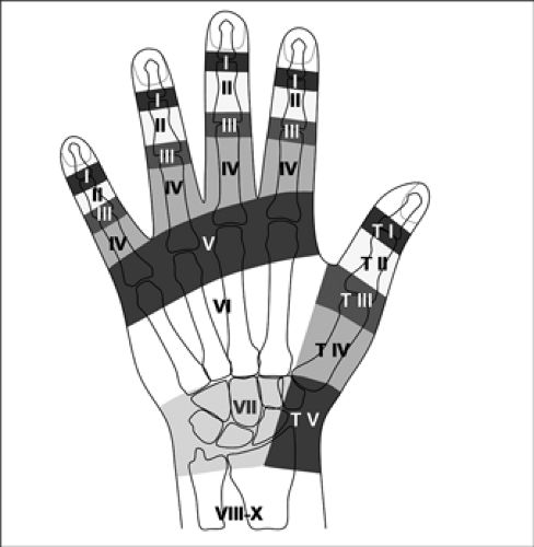
The Verdan classification of the finger extensor tendons. The anatomical distribution of extensor tendon regions: extrinsic extensors (regions VIII-X), wrist extensor region (region VII), dorsal hand (region VI), metacarpophalangeal (MCP) region (region V), proximal phalanx (region IV), proximal interphalangeal region (region III), middle phalanx (region II), and distal interphalangeal region (region I). The topography classification of the thumb is also noted: wrist extensor compartment (region V), metacarpal (region IV), MCP region (region III), proximal phalanx (region II), and interphalangeal region (region I).

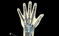
Dorsal forearm extensors. The dorsal forearm extensors consist of two groups. The superficial group comprises five muscles: (1) extensor carpi radialis longus; (2) extensor carpi radialis brevis; (3) extensor digitorum; (4) extensor digiti minimi; (5) extensor carpi ulnaris. The deep group includes: (6) abductor pollicis longus; (7) extensor pollicis brevis; (8) extensor pollicis longus; (9) extensor indicis.
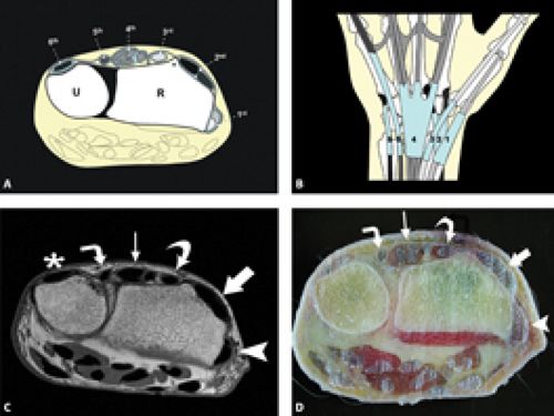
Wrist extensor compartments. A-B: The figure shows, (D) specimen photograph, and (C) T1-weighted MR images at the level of the wrist joint showing axial plane sections of the six extensor compartments: (1) abductor pollicis longus and extensor pollicis brevis (arrows); (2) extensor carpi radialis longus and brevis tendons (thick arrows); (3) extensor pollicis longus tendon (curved arrow); (4) extensor digitorum and deep extensor tendons (thin arrows); (5) extensor digiti minimi tendon (right angle curved arrow); (6) extensor carpi ulnaris tendon (asterisk).
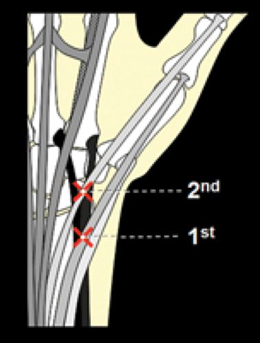
Crossing points of wrist extensor compartments. The illustration of the wrist extensor compartment shows the spatial relationships among the six extensor compartments. The first compartment tendon (abductor pollicis longus and extensor pollicis brevis) crosses the second compartment tendon (extensor carpi radialis longus and brevis) near the radial styloid, forming the first crossing point of the wrist tendons. The third compartment tendon (extensor pollicis longus) similarly crosses the tendon of the second compartment, forming the second crossing point at the wrist.
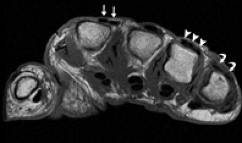
Diversity of extensor tendons. Axial T1-weighted magnetic resonance images show double extensor tendons (straight arrow) at the level of the metacarpophalangeal joints, triple extensor tendons of the ring finger (arrow), and double extensor tendons of the little finger (curved arrow). These are common variations and should not be misinterpreted as tendon pathologies.
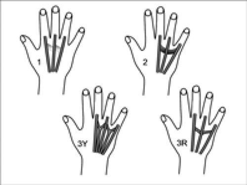
Tendon connections: The connective tissue of the tendons is a narrow band of connective tissue that extends between the extensors, redistributing forces, coordinating extension, and stabilizing the metacarpophalangeal joints. Several different types are described. Type 1 is a filamentous connection between the extensor digitorum and the long and ring fingers. Type 2 has morphological features between types 1 and 3, identified between the extensors of the middle and ring fingers, as well as between the ring and little finger tendons; Type 3Y is a tendon bundle representing a split extensor tendon, typically found between the little and ring finger tendons, and between the long and ring fingers. Type 3R is a more oblique tendon dislocation than type 3Y, usually found between the little and ring finger tendons and between the middle finger and ring finger.
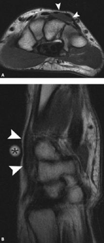
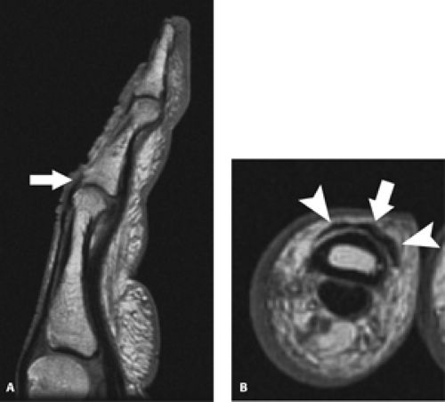
Short extensor tendon appears as a painless mass in patients without a history of trauma. T1-weighted (a) axial and (b) sagittal MR images of the hand show a well-defined oval area of moderate signal intensity (arrow), with the visible mass indicated below (asterisk). The signal intensity characteristics, morphology, and location of the dorsal wrist are consistent with short extensor tendon abnormalities.
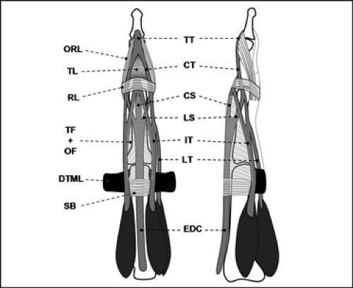
At the level of the metacarpophalangeal joints, the extensor tendons are composed of a soft tissue network (also known as the dorsal tendon sheath) that extends along the dorsal side of the fingers, stabilizing them and limiting their proximal migration. The extensor has various soft tissue components: EDC (extensor digitorum communis); SB (sagittal band); DTML (deep transverse metacarpal ligament); TF+ (intermetacarpal transverse fibers and oblique fibers); RL (oblique ligament (oblique support band)); TL (triangular ligament); ORL (oblique disc ligament); TT (terminal tendon (insertion point of the dorsal tendon sheath)); CT (central tendon); CS (central tendon bundle); LS, lateral bundle; IT, interosseous tendon; LT, lumbrical tendon.
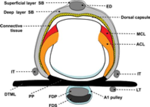
Sagittal band. The sagittal band (SB) refers to the fibrous sheets surrounding the joints (MCP) in both deep and superficial layers, fixed and stabilizing the toe extensor tendons (ED). It has a palmar attachment point at the palmar side (PP). This structure is unique to the MCP joint and closely related to surrounding soft tissue structures. MCL, medial collateral ligament; ACL, accessory ligament; IT, interosseous tendon; LT, lumbrical tendon; FDP, flexor digitorum profundus tendon; FDS, flexor digitorum superficialis tendon.
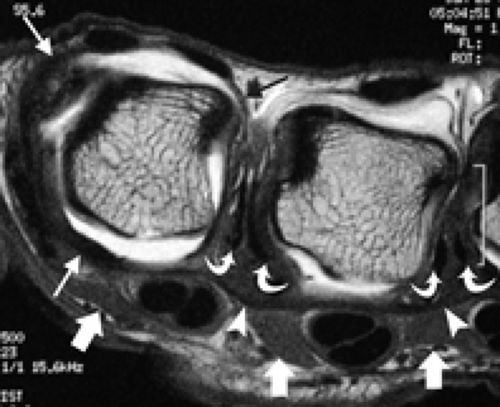
Anatomy of the metacarpophalangeal (MCP) joint. T1-weighted axial MR images after injecting contrast material into the MCP joint show the complex soft tissue anatomy surrounding the joint. The sagittal band surrounding the MCP joint is identified (thin arrows). The interosseous tendon observed dorsally (curved arrows) is located in the deep transverse metacarpal ligament (DTML) (arrow), while the palmar lumbrical muscles (thick arrows) can be identified as DTML.
Anatomy of the extensor tendons at the proximal interphalangeal joints. T1-weighted sagittal (a) and axial (b) MR images show the central bundle attaching to the middle phalanx and the lateral bundles (arrows) of the extensor tendons.
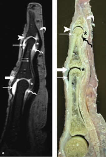
Anatomy of the extensor tendons in a cadaver specimen, T1-weighted fat-suppressed sagittal MR images after injecting contrast material into the proximal interphalangeal (PIP) and distal interphalangeal (DIP) joints. (b) The corresponding specimen photograph section shows the relationship between the extensor tendons and the joint capsule. The central sliding attachment at the base of the middle phalanx (thick straight arrow) and the terminal tendon attachment at the base of the distal phalanx (arrow) have been identified. The dorsal recesses of the PIP and DIP (asterisk) and the palmar recess of the joint (wavy arrow) are outlined.
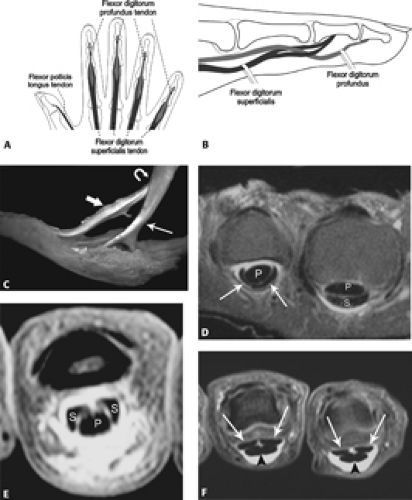
Anatomy of the flexor tendons. The diagrams (a-b) show the relationships and attachment sites of the flexor tendons. The anatomical photograph in the cadaver specimen (C) shows the flexor digitorum profundus (thick arrow) passing through (curved arrow) the flexor digitorum superficialis (thin straight arrow). A series of T1-weighted fat-suppressed MR tendonography images shows (D) the flexor digitorum profundus (P) located dorsally to the flexor digitorum superficialis (S) (thin straight arrow). The metacarpal, (E) flexor digitorum profundus (P) passes through the proximal phalanx superficial (S) split, and (F) the flexor tendons (arrow) palmarly to two separate parts at the level of the distal middle phalanx.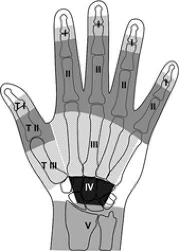
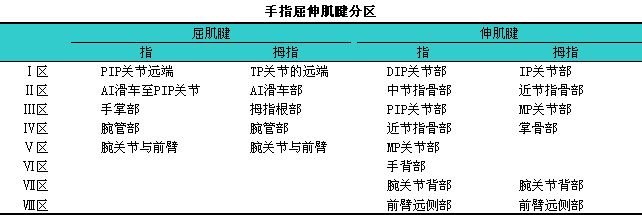
Regions of the flexor tendons. The image shows the five regions of the finger flexor tendons: Region I extends from the distal phalanx to the middle phalanx, Region II extends from the middle phalanx to the distal palmar crease, Region III extends from the metacarpophalangeal (MCP) level to the distal part of the flexor retinaculum, Region IV consists of the carpal tunnel, and Region V includes everything proximal to the carpal tunnel. In the thumb, Region I includes the interphalangeal joint area, Region II includes the MCP joint area, and Region III includes the metacarpal.
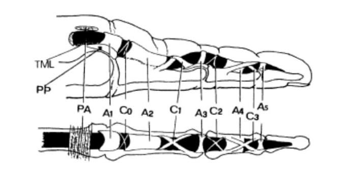
The pulley system of the finger flexor tendons.
The pulley system of the flexor tendons is a series of dense connective tissue bundles of varying widths, thicknesses, and shapes formed by the thickening of tendon sheaths at different sites, which restrain the flexor tendons and fully exert their flexion function.
The pulley system consists of 5 annular pulleys (A1, A2, A3, A4, A5), 4 cruciate pulleys (C0, C1, C2, C3), and 1 palmar aponeurosis pulley (PA), arranged in the order from proximal to distal as follows: PA, A1, C0, A2, C1, A3, C2, A4, C3, A5.
A1 pulley is located at the metacarpophalangeal joint, primarily attaching to the palmar plate of the MCP joint, with a small portion of fibers attaching distally to the base of the proximal phalanx and the lateral condyle.
A2 pulley is located at the proximal phalanx, attaching to the proximal 3/5 to 2/3 of the proximal phalanx, with fibers usually converging diagonally towards the lateral aspect of the base of the proximal phalanx.
A3 pulley is located at the proximal interphalangeal joint, attaching to the palmar plate of the proximal interphalangeal joint.
A4 pulley is located at the middle phalanx, attaching to the distal half of the upper segment of the middle phalanx on both sides.
A5 pulley attaches to the distal interphalangeal joint, anchoring to the palmar plate of the distal interphalangeal joint.
C0 pulley is located in the narrow area between A1 and A2, proximally attaching to the palmar plate of the MCP joint and distally attaching to the base of the proximal phalanx.
C1 pulley is located between A2 and A3, proximally attaching to the mid-portion of the proximal phalanx, crossing over the distal end of A2, and distally attaching to the palmar plate of the proximal interphalangeal joint, with some fibers joining the lateral bundle of the dorsal tendon sheath.
C2 pulley is located between A3 and A4, with the proximal end attaching to the palmar plate of the proximal interphalangeal joint and the distal end attaching to the base of the middle phalanx.
C3 pulley is located between A4 and A5, with the proximal end attaching to the mid-portion or base of the middle phalanx, crossing over the lateral side of the A4 pulley, and distally attaching to the palmar plate of the distal interphalangeal joint and/or the base of the middle phalanx.
Width of the pulleys: On average, the A2 pulley is the widest at 16.8mm; followed by the PA pulley, A1 pulley, and A4 pulley, measuring 8.0mm, 7.1mm, and 6.3mm respectively, while A3 and A5 are narrower at about 3-4mm.
Annular pulleys: Mainly composed of transverse fibers, they can be categorized into bony pulleys and palmar pulleys based on their attachment sites. A2 and A4 pulleys primarily attach to the phalanges, classified as bony pulleys; A1, A3, and A5 primarily attach to the palmar plates, classified as palmar pulleys.
The A1 pulley is thicker proximally and gradually thins distally. The A1 pulley may split into two or three parts, and the presence of a gap may relate to better adaptation of the tendon during flexion. The A1 pulley may also connect with adjacent pulleys.
The A2 pulley is the widest, predominantly consisting of transverse fibers, with crossing fibers visible in the proximal portion, thinner proximally, and thickest distally (about 1mm), slightly concave towards the dorsal side as it follows the curvature of the proximal phalanx, closely adhering to the tendon. The distal end is free, with the synovial membrane folding from the inner side to the palmar side, forming a significant synovial protrusion.
The A3 pulley is generally weaker, with a wider attachment site and a narrower mid-section.
The A4 pulley consistently appears and is well-developed, with the thickest mid-section (about 1mm) and slightly thinner sides (about 0.5mm). The inner side has a longitudinal ridge corresponding to the longitudinal groove on the palmar side of the flexor tendon.
The A5 pulley is generally weaker and may split into two or three parts.
Cruciate pulleys: The cruciate pulleys are typically formed by two bundles of oblique fibers crossing at a certain angle. The C0 and C2 pulleys are poorly developed and relatively weak, often absent in most fingers. The C1 and C3 pulleys are better developed, with the proximal part of the C3 pulley usually covering the distal end of the A4 pulley, and the distal end often fusing with the A5 pulley.
Palmar aponeurosis pulley: The PA pulley is composed of transverse fibers of the palmar aponeurosis. Generally, it consists of several bundles of loosely connected transverse fibers, with fine longitudinal fibers interwoven between them; and vertical fibers extend deep from the sides of the flexor tendons to attach to the deep transverse metacarpal ligament. The distal end of the PA pulley is located proximally to the heads of the metacarpals, covering the proximal end of the synovial sheath. This pulley does not closely adhere to the tendons, with a relatively thick layer of synovial membrane in between, located deep within it.
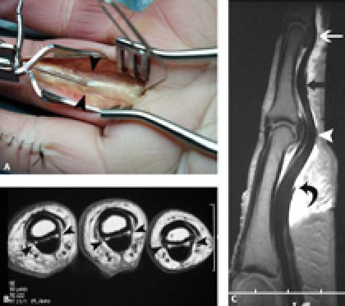
The pulley system of the finger flexor tendons. Cadaveric anatomy (A) shows the surgical probe beneath the A2 pulley of the flexor tendons (arrow). T1-weighted axial MR images of the proximal interphalangeal joint (B) demonstrate the normal morphology of the linear low signal A2 pulley (arrow) anchoring the flexor tendons to the adjacent bones. Cadaver specimens injected with contrast material show T1-weighted fat-suppressed sagittal MR images (C) with a well-defined A2 pulley (curved arrow), subtle thickening of the tendon sheath at the level of the proximal interphalangeal joint representing the A3 pulley (arrow), and a clear A4 pulley at the level of the middle phalanx (black straight arrow), with poor visibility of the A5 pulley at the distal interphalangeal joint (white straight arrow).
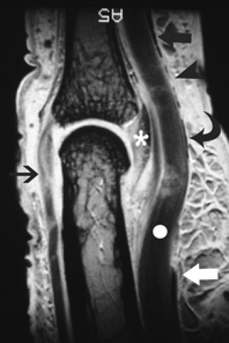
High-resolution sagittal MR images of the proximal interphalangeal joint obtained with a 1.5T local gradient coil reveal exquisite anatomical details, including the distal end of the A2 pulley (white thick arrow), A3 pulley (curved arrow), C2 pulley (arrow), and the A4 pulley (black thick arrow) of the flexor tendons (white circle). The palmar plate at the base of the middle phalanx (asterisk) and the central pulley attachment of the extensor tendons (thin black arrow) can also be observed.
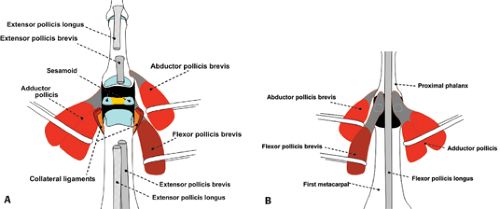
Anatomy of the thumb muscles and tendons. The diagrams of the dorsal (A) and palmar (B) aspects of the thumb illustrate the muscular and tendon anatomy concerning the bony and soft tissue structures.
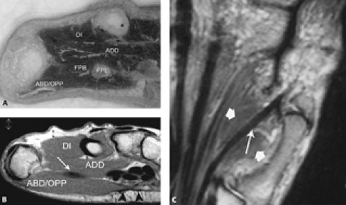
Anatomy of the thenar. Axial cadaver specimen photographs (A) and corresponding axial T1-weighted MR images (B) show the dorsal interosseous (DI), abductor pollicis (ADD), flexor pollicis longus (FPL), and tendon (white arrow), flexor pollicis brevis (FBP), abductor pollicis and adductor pollicis (ABD). In the MR image (B), the adductor pollicis can be seen in the central palmar fascia (arrow). Coronal proton density-weighted (C) MR images show the flexor pollicis longus tendon (long arrow) surrounded by the smaller belly of the flexor pollicis brevis (short arrow).
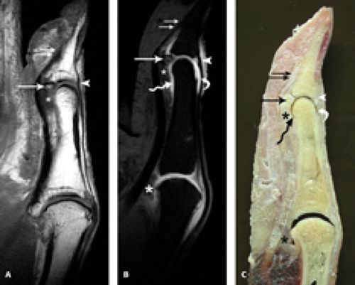
Anatomy of the tendons and joints of the thumb. T1-weighted sagittal MR images of the thumb in a cadaver specimen (A) allow identification of the distal clusters of flexors (double arrows) and extensors (arrow) and the insertion of the long flexor tendon of the thumb. Although not well-defined, the palmar plate (asterisk) of the interphalangeal (IP) joint and the adjacent sesamoid bone (arrow) can be observed. The corresponding T1-weighted fat-suppressed sagittal MR images after intra-articular injection of gadolinium at the MCP and IP joints (B) show the insertion sites of the flexor (double arrows) and extensor (arrow) long tendons of the thumb. The sesamoid bones of the IP joint (arrow), the palmar plate of the MCP and IP joints (asterisk), and the palmar (wavy arrow) and dorsal (curved arrow) recesses of the IP joint are indicated and better characterized as expanded joints. The specimen photograph (C) shows the overall appearance of the above structures.

Anatomy of the dorsal tendon sheath of the thumb. The extensor apparatus of the thumb consists of the tendons of the abductor pollicis longus, extensor pollicis brevis,
the dorsal tendon sheath (equivalent to the sagittal band), and a triangular expansion formed by oblique fibers released from the distal tendon of the adductor pollicis, along with a triangular expansion formed radially from the tendons of the flexor pollicis brevis and abductor pollicis.
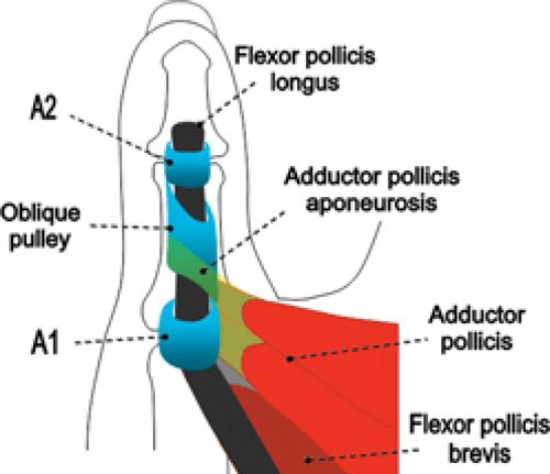
Anatomy of the pulley system of the thumb. The pulley system of the thumb includes two annular pulleys located at the level of the metacarpophalangeal joint (A1) and interphalangeal joint (A2). There is an inserted oblique pulley located at the proximal phalanx level, closely related to the attachment of the tendon sheath of the adductor pollicis.