Diffusion tensor imaging (DTI) is an emerging imaging expression method developed based on conventional magnetic resonance imaging technology. DTI is a special form of MRI that not only displays the signal intensity of each voxel but also calculates specific data on tensor orientation in three-dimensional space. Each voxel’s individual arrow is organized based on the differences in neural bundle orientation, generating a set of directional arrows. Through image data processing, each set of directional arrows is processed to form linear images of neural bundles.
The white matter fiber bundles in the brain are arranged geometrically. Since diffusion magnetic resonance imaging can clearly display the white matter fiber bundles, it allows for multi-angle studies of their arrangement. It is now possible to observe the anisotropy of water molecule movement within white matter fiber bundles in depth. The density of white matter bundle arrangement is proportional to the anisotropy of that area. In brain tissue, the anisotropy of white matter is higher compared to that of the cerebral cortex, which has a relatively lower anisotropy. Due to the presence of cell membranes, molecules must complete transmembrane movement during their movement between inside and outside the cells and tissues, during which water molecules will undergo diffusion phenomena.
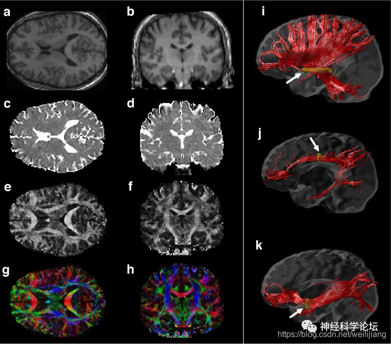
Due to the obstruction of the neural cell membrane, the movement or diffusion of water molecules along white matter fibers is faster than that perpendicular to the fibers, resulting in anisotropy. The differences in diffusion between parallel and perpendicular movements establish the imaging basis for diffusion tensor imaging. The fractional anisotropy (FA) value is expressed in the range of 0 to 1 and represents the ratio of the anisotropic component of water molecules to the entire diffusion tensor. A smaller value indicates less restricted diffusion, while a larger value indicates increased regularity and directionality of the tissue, thus enhancing neural conduction function. Therefore, FA values can be used to infer the degree of arrangement of cellular structures within brain white matter fiber bundles and the integrity of tissue structure.
DTI not only describes the directionality of water molecule diffusion within tissues through fractional anisotropy (FA) but also provides more specific descriptions of the direction and magnitude of water molecule diffusion through the apparent diffusion coefficient (ADC), mean diffusivity (MD), axial diffusivity (AD), and radial diffusivity (RD). The ADC value measures the diffusion movement of water molecules within tissues, reflecting the degree of displacement of water molecules in the diffusion-sensitive gradient direction. ADC is influenced by factors such as the temperature of water inside and outside cells, cell membrane permeability, viscosity, and proportion.
MD can reflect the total water content in tissues, expressing the overall diffusion activity and molecular replacement of water molecules; in the parallel axial position, AD describes the direction where the diffusion and movement of water molecules encounter the least resistance, which is displayed as the maximum value in the tensor. AD is more sensitive to the integrity and degeneration of axons. In the perpendicular axial position, RD is described, with the value depending on the average value calculated from two relatively low tensor values. RD can express the integrity of myelin. DTI has unique advantages in identifying and estimating neural bundles at the subcortical level and is widely used in both clinical diagnosis and research exploration, including studies on various brain diseases such as stroke, Alzheimer’s disease, and brain aging.
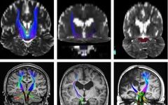
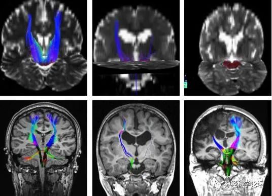
Diffusion is a vector with both magnitude and direction. In DTI, the displacement of water molecules within tissues is measured in at least six directions [3], whereas diffusion is determined in only one or three directions, which may lead to erroneous conclusions about tissue structure. DTI is a more advanced form of diffusion weighted imaging and is a new MR imaging technology based on DWI. It utilizes various parameters and data processing to reflect changes in diffusion within imaging voxels quantitatively and directionally, allowing for a directional and quantitative evaluation of the anisotropy of brain white matter. Additionally, DTI images can be displayed using specific post-processing software with principal feature vector maps, thereby showing the direction and integrity of white matter in the images. This provides the possibility to study the pathways of white matter and reveal the relationship between various brain lesions, including brain infarction and brain tumors, and white matter fiber pathways, demonstrating greater superiority and potential in displaying white matter fiber lesions. Fiber tractography is currently the only imaging technology that can provide the location and characteristics of human brain white matter fiber structures in vivo, non-invasively, and individualized. It can visually display the relationship between tumors and surrounding white matter fibers, thereby better guiding surgeries to maximize tumor tissue removal while protecting normal brain tissue.
More Exciting Reads
Exploring Difficulties | Essential Tremor Is More Than Just Tremors!
Clinical Management of Non-Motor Symptoms in Parkinson’s Disease (2020)
Exploring Difficulties | The Kaleidoscope in Neurological Diseases!
The Future of Behavioral Sleep Medicine
Complementary and Alternative Therapies (CAM) for Parkinson’s Disease