“Complementarity” and “Integration”
Multimodal imaging unveils the mysteries of life from all angles
1
Understanding Life: Multimodal Biomedical Imaging Technology
In recent years, rapidly developing biomedical imaging technologies can depict the structure and function of living organisms at different scales, such as in vivo, tissue, and molecular levels. However, single-modal representation is easily affected by objective factors and has certain limitations. Therefore, the complementarity and integration of various imaging modalities is a development trend in the field of biomedical imaging, making it possible to dynamically depict the processes of life activities across scales, which is of great significance.
Multimodal imaging can integrate various imaging paradigms such as optical, electrical, mechanical, magnetic, and radionuclide imaging, achieving cross-scale and multimodal imaging from molecular to living levels, from angstroms to meters. Multimodal biomedical imaging technology provides research tools and means for complex life science issues and major diseases, allowing for comprehensive visualization and precise measurement of the overall structure, dynamic physiology, and metabolic processes of living organisms, thereby revealing the mysteries of life and disease.
2
Multimodal Imaging Solving Problems in Life Medicine Research
Multimodal imaging leverages the advantages of various imaging technologies for “complementarity” and “integration,” breaking through the scale limitations of single-modal imaging, and features cross-scale, all-dimensional, and visualization characteristics. It is widely used in cutting-edge life medicine disciplines such as oncology, brain science, and cardiovascular research. Below, we will take tumor research as an example to introduce the application of multimodal imaging technology in this field.
In Sight • In Vivo Imaging
In vivo imaging allows for real-time observation and dynamic tracking of physiological responses and pathological processes within living organisms by tracking a group of living tissues or small animals at different time points, facilitating studies on cellular activities and gene behaviors, which helps to better understand the occurrence and development patterns of human diseases and research prevention and treatment measures. Currently, commonly used in vivo imaging technologies include magnetic resonance, ultrasound or photoacoustic imaging, radioactive nuclide imaging, chemiluminescence or fluorescence imaging, etc. In vivo imaging is not only applied clinically but also plays a significant role in preclinical research.
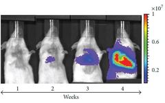
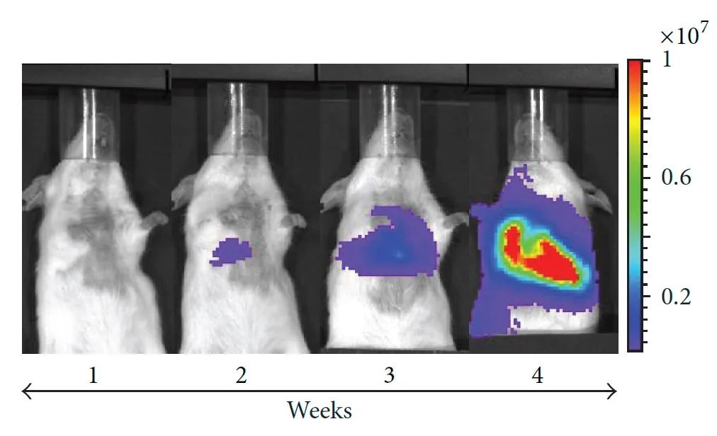
In tumor research, in vivo imaging technology can visually detect and evaluate the growth, metastasis of tumors within animal bodies, as well as the distribution and efficacy of tumor-related drugs. The metastasis of tumor cells is a major cause of cancer mortality. Early detection of cancer at primary and metastatic sites through non-invasive techniques is crucial for disease treatment intervention and management. By providing complementary information through chemiluminescence, radionuclide labeling, and magnetic resonance imaging, it is possible to monitor the metastasis of tumor cells within the body visually and accurately measure tumor development.
Dynamic monitoring of the development and metastasis of luciferase-labeled melanoma cells in vivo through chemiluminescence imaging
PET/CT and MRI accurately locate tumor metastases
Micro Insights • Tissue Imaging
Accurate early diagnosis of cancer is the basis for effective tumor treatment, where imaging of tumor tissues is a critical component of cancer research and clinical diagnosis, holding significant biological and clinical importance.
Current mainstream pathological tissue imaging methods primarily use optical imaging, which is easily affected by strong optical backgrounds, unstable signals, inaccurate quantification, and incompatibility among different optical methods, thus impacting the accuracy of tissue pathological detection. Electron microscopy overcomes the low resolution limitation of optical microscopy, enabling detailed observation of the ultrastructure of tumor tissues, finding differentiation markers of tissue cells, confirming and distinguishing corresponding tumor types, and holding an irreplaceable position in modern tumor research and diagnosis. The multimodal imaging technology that combines electron microscopy and optical imaging continuously drives tumor research, providing important scientific basis for tumor biological characteristics, evolution patterns, disease diagnosis, and treatment monitoring.
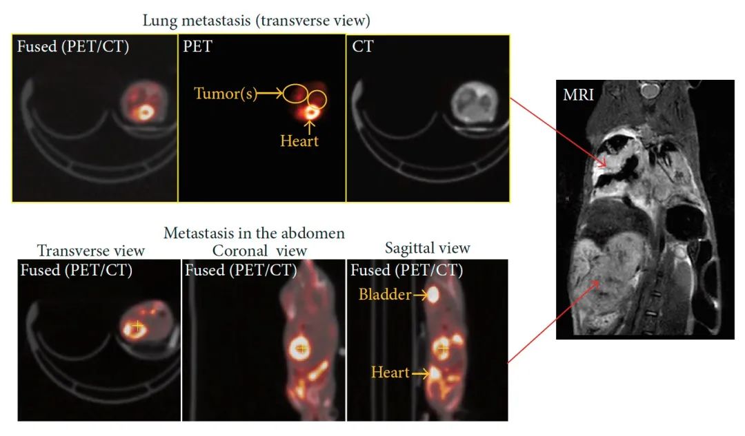
Research published on the cover of Science in April 2022 utilized low-temperature imaging technology and focused ion beam scanning electron microscopy to observe the interaction between cell tissues and the tumor microenvironment, vividly showcasing the process of T cells “battling” cancer cells, providing important scientific evidence for studying tumor evolution and the immune system’s role in combating cancer cells.
The process of T cells “battling” cancer cells
Decoding the Code • Molecular Imaging
The rapid development of cell biology and molecular biology has greatly deepened our understanding and knowledge of tumors. In-depth studies of oncogenes, tumor suppressor genes, cell cycle, drug resistance-related genes and proteins, cellular signal transduction systems, and even the Human Genome Project have made it possible to observe and understand tumors from different molecular levels. Using some precise imaging methods, we can directly “see” the changes in these biomolecules, thus providing intuitive and effective criteria for determining tumor stages, evaluating treatment efficacy, and other research issues.
The following image uses laser confocal microscopy to observe the expression and colocalization of autophagosomal and lysosomal membrane proteins in prostate cancer tissue samples, reflecting the malignancy of cancer cells and determining whether the patient is more suitable for surgical treatment or radiotherapy, which plays an important role in clinical diagnosis and treatment guidance.
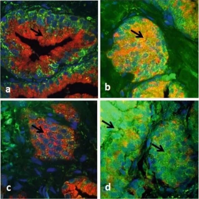
Immunofluorescence imaging of prostate cancer tissue samples
(Green represents autophagosomal membrane protein LC3A, red represents lysosomal membrane protein LAMP2a, yellow represents expression and colocalization)
BRCA1-BARD1 tumor suppressor factor is an E3 ubiquitin ligase essential for repairing DNA double-strand breaks, which is significant for DNA repair. To understand its mutation and relationship with cancer, scientists used cryo-electron microscopy to perform three-dimensional analysis of its molecular structure, revealing important functional domains of BRCA1-BARD1, which has significant implications for cancer prevention and drug development.
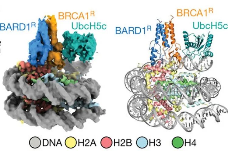
Cryo-electron microscopy analysis of the structure of BRCA1R–BARD1R–UbcH5c–NCP complex
The continuous development of multimodal imaging technology will continue to promote preclinical oncology research, providing important evidence for tumor evolution processes at in vivo, tissue, and molecular levels, achieving high-resolution longitudinal studies, and holding immense application potential in biomedical research and molecular diagnosis of diseases.
3
A Powerful Tool for National Translational Medicine Facilities
The “National Major Scientific and Technological Infrastructure for Translational Medicine (Shanghai)” project is one of the key areas in the national planning of the 12th Five-Year Plan. The construction goal is translational research on three major diseases: tumors, metabolic diseases, and cardiovascular diseases; verification of the effectiveness of drugs, reagents, and materials; and the transformation of key common technologies for large-scale high-end medical equipment.
The Biomedical Imaging Technology Center is an important component of the National Major Scientific and Technological Infrastructure for Translational Medicine (Shanghai), focusing on biomedical imaging and preclinical animal (molecular) imaging research, covering research fields such as oncology, brain science, cardiovascular, and pharmacokinetics, and has laid out imaging technologies such as magnetic resonance, ultrasound, photoacoustic, chemiluminescence/fluorescence imaging, radioactive nuclide labeling, confocal, scanning electron microscopy, transmission electron microscopy, and cryo-electron microscopy.
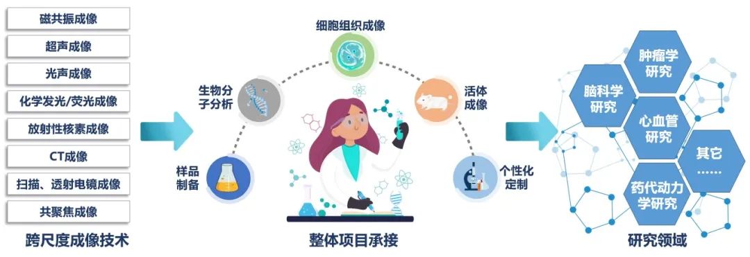
One-stop imaging facility cluster of the Biomedical Imaging Technology Center
The Biomedical Imaging Technology Center integrates multiple biomedical imaging technologies organically, establishing an advanced biomedical imaging facility cluster that is large-scale, multidisciplinary, goal-oriented, and multimodal. It can provide a full-process, one-stop imaging technology support from molecular to living, from experimental design to data analysis, providing a full-function research platform for basic biomedical research, medical innovation, and achievement transformation in China, continuously promoting the development and progress of the life sciences field.
Contact: Cao Jingjing
Email: [email protected]
References:
1. Adiseshaiah, Pavan P., et al. Longitudinal Imaging of Cancer Cell Metastases in Two Preclinical Models: A Correlation of Noninvasive Imaging to Histopathology[J].
International Journal of Molecular Imaging, 2014, 2014(102702): 1-13.
2. Ritter A T, Shtengel G, Xu C S, et al. ESCRT-mediated membrane repair protects tumor-derived cells against T cell attack[J]. Science, 2022, 376(6591): 377-382.
3. Kalamida D, Giatromanolaki A, Koukourakis M I. Autophagy: Characterization of the “Autophagic Flux” in Prostate Cancer Tissue Biopsies by LC3A/LAMP2a Immunofluorescence and Confocal Microscopy[M]. New York, NY: Humana Press, 2019: 555-560.
4. Hu Q, Botuyan M V, Zhao D, et al. Mechanisms of BRCA1–BARD1 nucleosome recognition and ubiquitylation[J]. Nature, 2021, 596(7872): 438-443.