Dear teachers and friends, it’s time for another session on the basic operations and parameters of magnetic resonance imaging. In previous articles, we introduced some meanings of the Philips MRI operating interface, the names and significance of scanning sequences, the operation of Geometric parameters, and the meaning of Contrast. In this issue, we will continue to explain the significance and usage of parameters under the Contrast parameter section.Previous article links:Philips MRI Operations: Scanning CardPhilips MRI Operations: Parameter Information and Data Management (1)Philips MRI Operations: Parameter Summary and GeometryPhilips MRI Operations: Parameters ContrastPhilips MRI Operations: Parameters Contrast (2)This issue continues to introduce the main parameter contents in the contrast comparison parameter section, focusing on diffusion-weighted imaging, i.e., DWI.
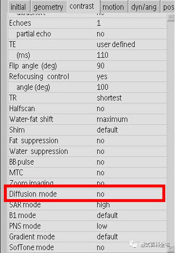
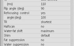
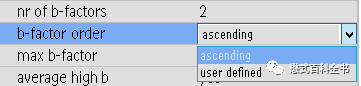
Figure 1:Parameter settings related to DWI in the Contrast parameter section
Diffusion can be understood as diffusion or spreading, mainly referring to the Brownian motion of molecules (MR primarily refers to water molecules) at a certain temperature. Most textbooks translate DWI as diffusion-weighted imaging, although some books translate it as diffusion-weighted imaging.
In fact, diffusion and spreading mean similar things. Diffusion reflects the result and carries a sense of “isotropy”; while spreading emphasizes the process and carries a sense of “anisotropy”.
Therefore, most domestic books translate DWI as diffusion-weighted imaging; when translating DTI, they translate it as diffusion tensor imaging.
Related articles on DWI can be found in the links below:
How to Effectively Perform DWI Sequences and DWI Scanning Parameter Techniques
Overview of Extended Applications and Various Advanced Diffusion Models of DWI
Why Not Only Look at DWI Images to Judge Diffusion Restrictions, But Also Consider ADC Images
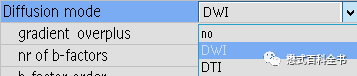
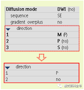
Figure 2: Diffusion mode
First, select Diffusion Mode to activate the DWI-related scanning sequence. There are three options:namelyno,DWI, andDTI.nomeans that this option is not selected, i.e., the diffusion-sensitive gradient is not activated;DWIis a technique primarily used to detect the movement direction and position of water molecules, suitable for examinations of various body parts and similarPETexaminations;DTIis a technique used to describe the diffusion characteristics of water molecules, currently mainly used for tracing white matter fiber bundles.
Previous article link: Whole-body MRI Technology (1)
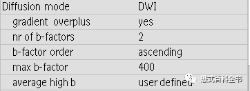
Figures 3 and 4:Related parameter settings for Diffusion sequencesIn DWI mode, we can set the number ofB factors innr of b-factors. In Philips standard mode, up to 16 B values can be scanned simultaneously in one sequence; the research version allows for even more. Once we set the number ofB values, we then need to set the specificB value sizes. Inb-factor order, we can choose the order ofB values (whether to increase sequentially or user-defined). If we choose user-defined, we can input the desired values, such as 0, 1, 2… Moreover, in the Philips system, the minimum step size for changing B values is 1. For example, if we set 5 B values as 0, 1, 2, 3, 4. However, we generally do not set such dense B value changes. For instance, in IVIM, we typically set 5 to 10 B values, starting from small to large, where 200 is a turning point; we can set more densely before that and more sparsely afterward, such as: 0, 10, 20, 50, 80, 100, 200, 500, 800, 1000.
If set to Ascending, it indicates an increase. As shown in the figure above, if we set the maximumB value to 1000, then according to the previously setB value count of 2, it will automatically generate twoB values within the range of 0-1000, namely 0 and 1000.
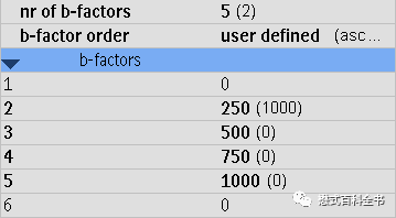
Figure 5: User-defined number of B values and size of each B value
Choosinguser definedallows us to customize the desiredB values. As shown in the figure above, if we setnr of b-factors to5, we can then set our desiredB values inb-factor order. What does the B value represent?B value is thediffusion sensitivity factor, with units ofs/mm², which is an indicator of the ability to detect diffusion motion.B value increases indicate greater sensitivity to the diffusion motion of water molecules. Of course, theoretically, asB value increases, the signal-to-noise ratio (SNR) of the image decreases, which is easy to understand, because asB value increases, the applied gradient becomes larger, and the duration of the gradient becomes longer, which results in a longerTE time and a lower SNR of the image.
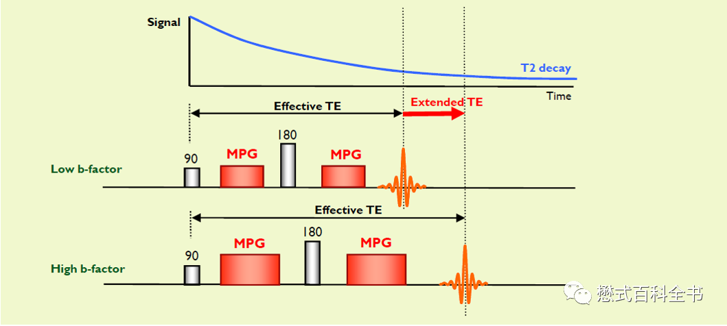
Figure 6: A larger B value indicates a greater applied diffusion motion-sensitive gradient, generally leading to extended TE time and decreased SNR

Figure 7: Assigning corresponding excitation counts to each B value
SinceB value increases lead to lower image SNR, we can improve the SNR by increasing the excitation count (NSA). In Philips MR, we can selectuser defined underaverage high b, where we can assign a correspondingNSA for eachB value.
This approach has the advantage of reasonably distributing NSA to different B values, as high B value images experience a decrease in SNR, while low B value images do not decrease significantly. Therefore, it is unnecessary to apply high excitation counts to every B value, as this would significantly extend scanning time.
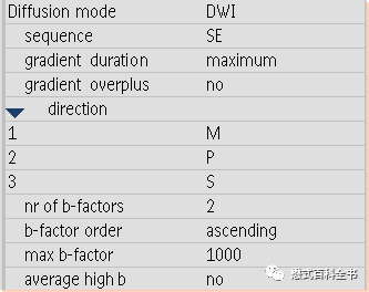
Figure 8: Directions of applied diffusion motion-sensitive gradients
In performing DWI sequences, we apply diffusion motion-sensitive gradients in one or more directions.
If we select no for gradient overplus, we need to define the directions in which the diffusion motion-sensitive gradients are applied. We can choose to activate three directions simultaneously, or two, or even one, with various combinations of different directions. Here, M represents the readout gradient direction (frequency encoding direction) Measurement; P represents the phase gradient direction (i.e., phase encoding direction) Phase; S represents the slice gradient direction (i.e., slice encoding direction) Slice.
Generally, when we perform isotropic DWI scans, we activate all three directions simultaneously to ensure that the influence on water molecules is consistent in all directions. However, in certain cases, such as nerve root imaging, we can activate only one direction based on the nerve trajectory to detect nerve root morphology through anisotropy.
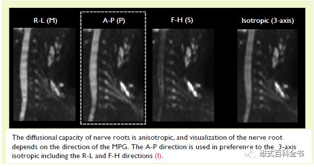
Figures 9 and 10:During brachial plexus nerve root imaging, diffusion motion-sensitive gradients are applied only in the anterior-posterior direction for the best visualization of the nerve roots
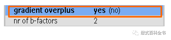
Figure 11: Usage of gradient overplus
As shown in the figure above, if the user selects gradient overplus, the system automatically applies diffusion motion-sensitive gradients in three directions and achieves reduced TE values through combinations.
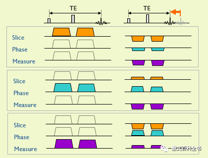
Figure 12: Effect after using gradient overplus (Image selected from Japanese team training materials)
As shown above, since it is necessary to activate diffusion motion-sensitive gradients in three directions, using OG can achieve a reasonable combination of gradients to reduce TE time, thus improving SNR.
Many factories of Philips have a DWI sequence named DWI_og, and many customers ask what this og means; in fact, this og refers to the DWI sequence that uses Gradient Overplus, which should clarify things.
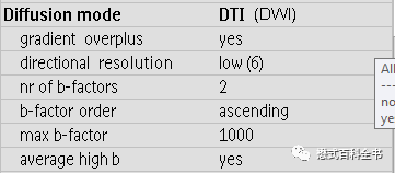
Figure 13: DTI mode
If the Diffusion mode is set to DTI instead of DWI, we can activate DTI-related sequence scanning using diffusion tensor imaging.
A tensor is an engineering concept; the DWI scans we mentioned earlier are primarily based on an isotropic model, where the probability of water molecules moving in all directions is the same; while DTI is based on an anisotropic model.
So what are the differences between DTI and DWI in terms of sequences?
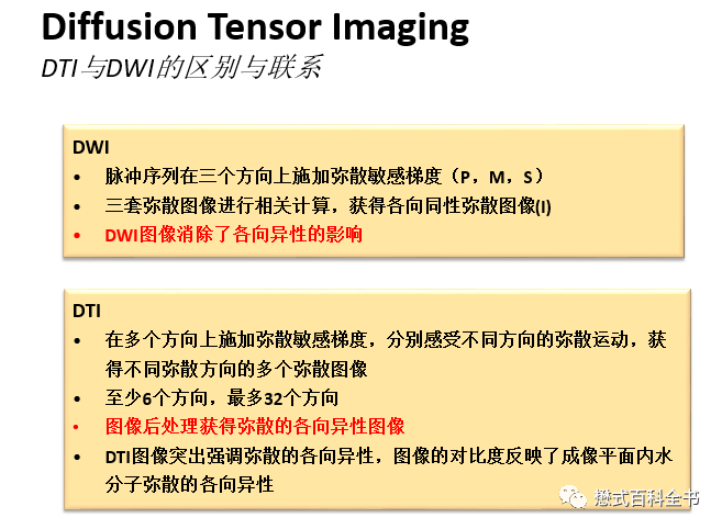
Figure 14: Similarities and differences between DTI and DWI sequences
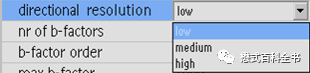
Figure 15:Direction of DTI
In DTI sequences, an important parameter is directional resolution, which refers to the number of applied diffusion motion-sensitive gradient directions.
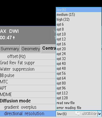
Figure 16:Different DTI sequences have different directional resolutions, reflected in the number of directions
Different DTI sequences have different resolutions for fiber bundles due to the varying directions of the diffusion motion-sensitive gradients applied in DTI.
The more directions, the higher the DTI directional resolution, but the longer the scanning time; conversely, the fewer directions, the lower the resolution but shorter the scanning time.
If we want to perform DTI, we need to apply at least 6 directions, which is indicated as low (low direction count 6) in the parameters.
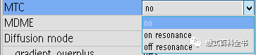

Figure 17: MTC-related parameters
MTC stands for magnetization transfer contrast or magnetization transfer imaging technology. Using this parameter activates magnetization transfer. For specific content, see previous articles:
What is Magnetization Transfer MTC?
This concludes the content of this issue. Thank you all for your support.Thank you for your supportYour appreciation is encouragement for our work

2019.08.21 in Shanghai Leo and Viktor
Follow the Encyclopedia of Mao, for more exciting original content.
Welcome to subscribe to my WeChat Official Account (personal blog).
Добро пожаловать в мой блог WeChat.

Copyright Statement:
Copyright belongs to “Encyclopedia of Mao”. Personal collection, forwarding, and sharing are welcome. Any other media, websites, public accounts, platforms, or forums that need to reprint or quote must obtain authorization. Without authorization, theft is prohibited! After obtaining authorization, it must be prominently indicated “Reprinted from: Encyclopedia of Mao”, and a link to the original article must be set in the reading original section, allowing readers to return to my original article. Thank you for your support! Sincerely, the author.