
Every day we need to select information from different positions in space. Since the total energy available to the brain in a short time is limited, if the brain wants to filter more spatial information, it needs to quickly and efficiently allocate attention resources. This process is accompanied not only by effective representation and encoding of information, but also by flexible allocation of attention based on past experiences or prior information.
On January 25, 2022, the Song Yan research group at the State Key Laboratory of Cognitive Neuroscience and Learning, Beijing Normal University, published an article titled “The neurovascular couplings between electrophysiological and hemodynamic activities in anticipatory selective attention” in the internationally renowned cognitive neuroscience journal Cerebral Cortex. The study utilized simultaneous recording and fusion analysis of EEG and fNIRS to map the activation response patterns of attention with high spatial and temporal resolution, further revealing the interesting neurovascular coupling mechanism behind attention. We focus our attention on visual stimuli that are most relevant to the current goal while ignoring inputs that are irrelevant to the behavior. These two processes are referred to as target enhancement and interference suppression. However, in previous studies, the suppression of interference was often tightly bound to target enhancement. This study innovatively combined target and interference probability distributions with spatial information (Figure 1), successfully achieving the separation of the target enhancement and interference suppression processes.
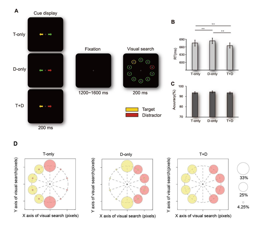
Figure1 Experimental Design and Behavioral Results
From EEG data of normal adults, researchers found that target enhancement induces an early positive alpha (8 ~ 12 Hz) energy lateralization modulation in the posterior brain (Figure 2A), while interference suppression induces a later negative alpha lateralization modulation (Figure 2B). From fNIRS data, it was found that the expectation of targets and distractors induces similar lateralized activities of oxygenated hemoglobin (HbO) in the lateral striatum region (Figure 2 D,E), but the activation of the visual cortex for both is significantly different.
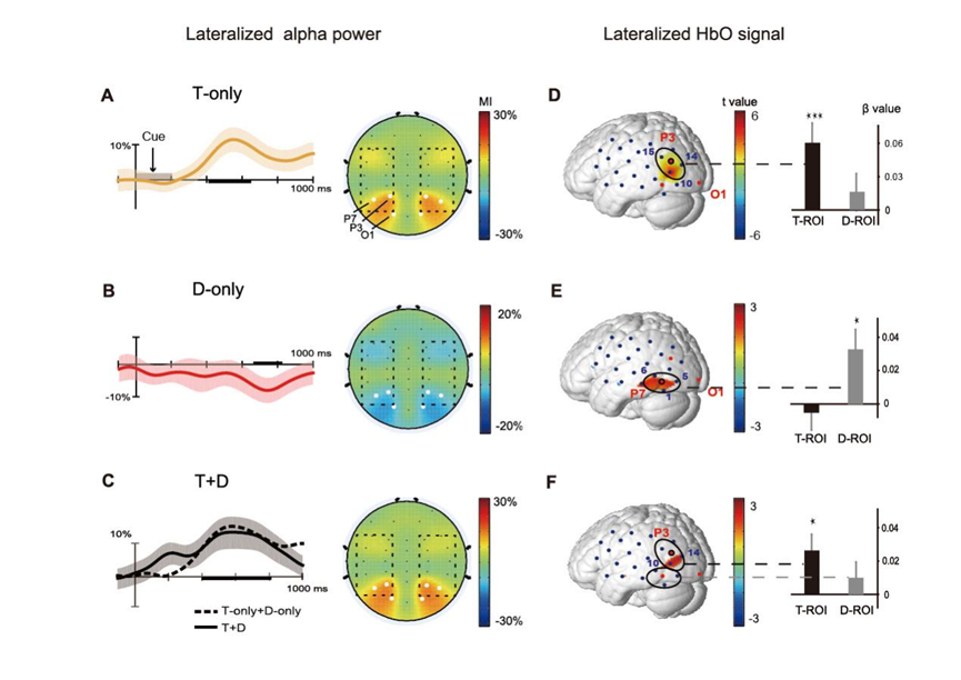
Figure 2 Three different electrophysiological neural responses (left) and hemodynamic signal responses (right) induced by three different cues
More importantly, the researchers discovered a specific neurovascular coupling relationship between alpha activity and HbO signals. This relationship manifests as a positive coupling effect during target enhancement (Figure 3A) and a negative coupling effect during interference suppression (Figure 3B). Thus, the researchers found multiple separations in time, space, and coupling relationships for target enhancement and interference suppression in attention. Interestingly, the interference suppression-related alpha activity exhibited a negative modulation pattern. This pattern ensures that attention resources are less attracted by subsequent distractors.
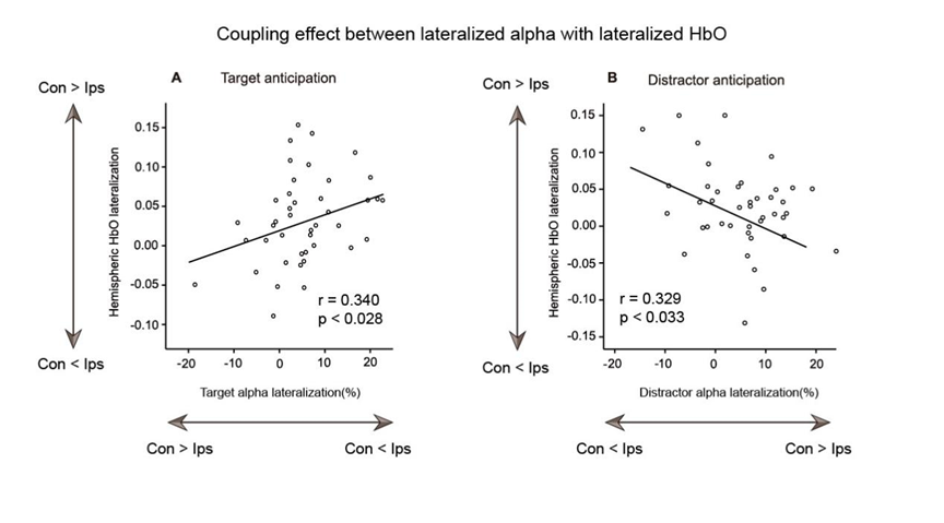
Figure 3 Opposite neurovascular coupling relationships for target enhancement (A) and interference suppression (B)
However, the above analysis only qualitatively examined the neurovascular coupling relationships, and quantifying these relationships remains a problem to be solved. Since the brain’s electrical activity and hemodynamic activity provide “orthogonal” neurophysiological information from two aspects, there is no perfect spatiotemporal correspondence between the two. This increases the difficulty of conducting fusion analysis. To address this issue, the researchers first employed an alpha-informed analysis method in the analysis (Figure 4). They extracted alpha information from individual trials to analyze the variability of EEG signals across trials and modeled it against continuous HbO signals, thereby achieving quantitative analysis of the coupling relationship.
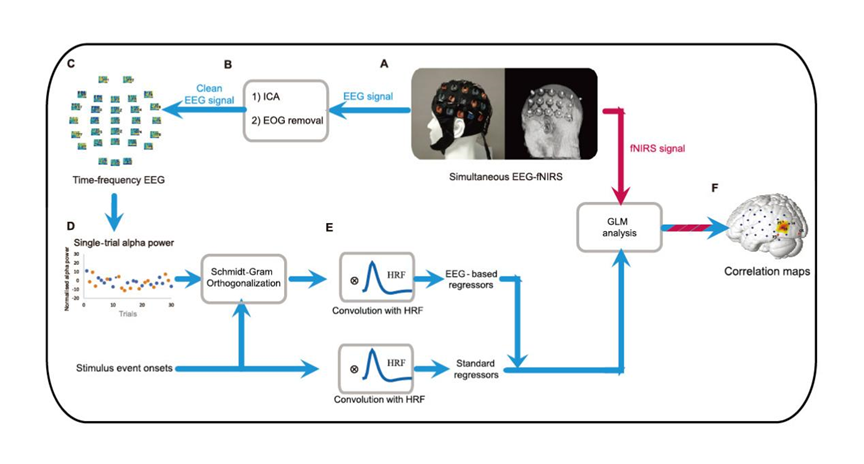
Figure 4 Flowchart of alpha-informed analysis
The results not only further validated the directional differences in neurovascular coupling (Figure 5 ABC), but also found that different coupling strengths seem to have a significant impact on final behavioral responses. This is reflected in individuals with better behavioral performance being able to benefit more effectively from positive alpha-HbO coupling during target enhancement, or utilizing negative alpha-HbO coupling more effectively to suppress upcoming distractors during interference suppression (Figure 5D).
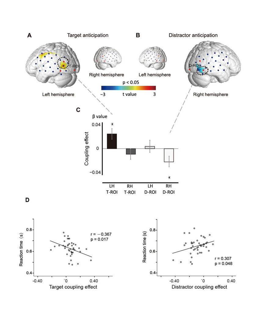
Figure 5 Relationships between lateralized alpha energy and raw blood oxygen activity during target enhancement (A) and interference suppression (B), (D) Quantified neurovascular coupling strength can explain variations in behavioral response times.
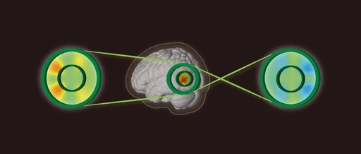
The above results not only provide new evidence for the separation of mechanisms in target enhancement and interference suppression in attention from the perspective of neurovascular coupling, but also help researchers establish two neurovascular coupling models to explain the division of labor and cooperation exhibited by neuronal electrophysiological activity and hemodynamic signals during the attention process. The researchers vividly used the “conveyor belt-driven model” to explain the neurovascular coupling mechanisms for target enhancement (Figure 6A: open conveyor belt) and interference suppression (Figure 6B: cross conveyor belt).
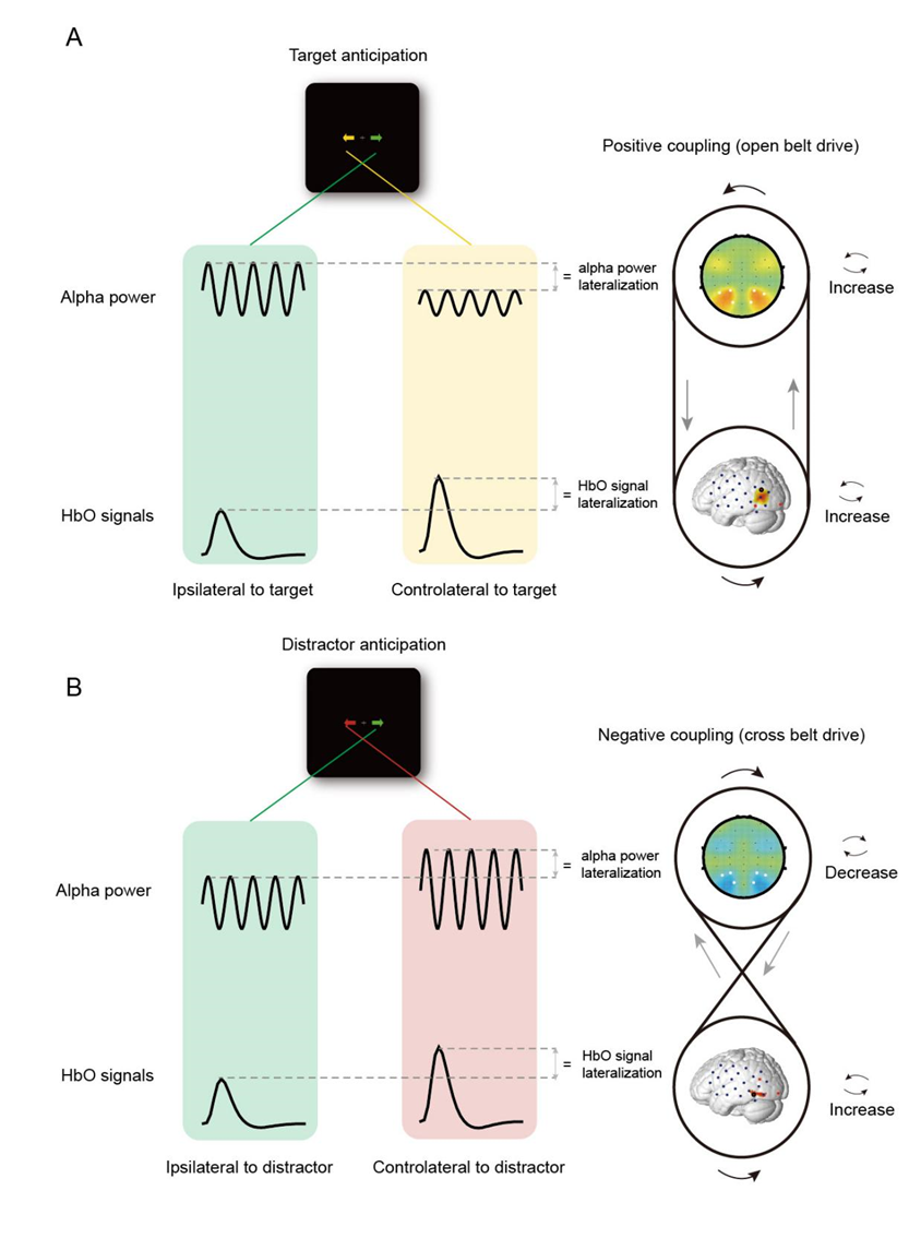
Figure 6 Neurovascular coupling mechanisms in different attention processes as “conveyor belt” models. A Open conveyor belt driving model for target enhancement B Cross conveyor belt driving model for interference suppression.
It is worth noting that this study utilized a non-invasive, mutually non-interfering, minimally motion-sensitive, and ecologically valid method of simultaneous fNIRS–EEG recording, providing new ideas for research on cognitive neuroscience effects. It also offers new ideas and methods for future research on attention mechanisms in children with attention difficulties and other disorders.
This study is an extension and continuation of the Song Yan research group’s exploration of the neurovascular coupling mechanisms in attention processes in recent years (Huang et al., NeuroImage 2015; Zhao et al., Human Brain Mapping 2019) . The first author of this paper is Zhao Chengguang, a postdoctoral researcher at the Zhuhai campus of Beijing Normal University, with Li Dongwei and Guo Jialiang as the second and third authors, and Professor Song Yan as the corresponding author. Thanks to Professor Li Xiaoli from Beijing Normal University, Professor Liu Hanli from the University of Texas at Arlington, and Professor Ding Yulong from South China Normal University for their guidance and assistance in this research. This research was funded by the National Natural Science Foundation, the Open Project of the National Key Laboratory, and the Basic Scientific Research Program of China’s National Defense.
Paper link: https://doi.org/10.1093/cercor/bhab525
References:Zhao C, Guo J, Li D, Tao Y, Ding Y, Liu H, Song Y. 2019. Anticipatory alpha oscillation predicts attentional selection and hemodynamic response. Human Brain Mapping. 40:3606–3619.
Huang J, Wang F, Ding Y, Niu H, Tian F, Liu H, Song Y. 2015. Predicting N2pc from anticipatory HbO activity during sustained visuospatial attention: a concurrent fNIRS-ERP study. NeuroImage. 113:225–234.
———–———-———-———-——
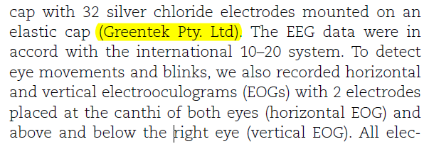
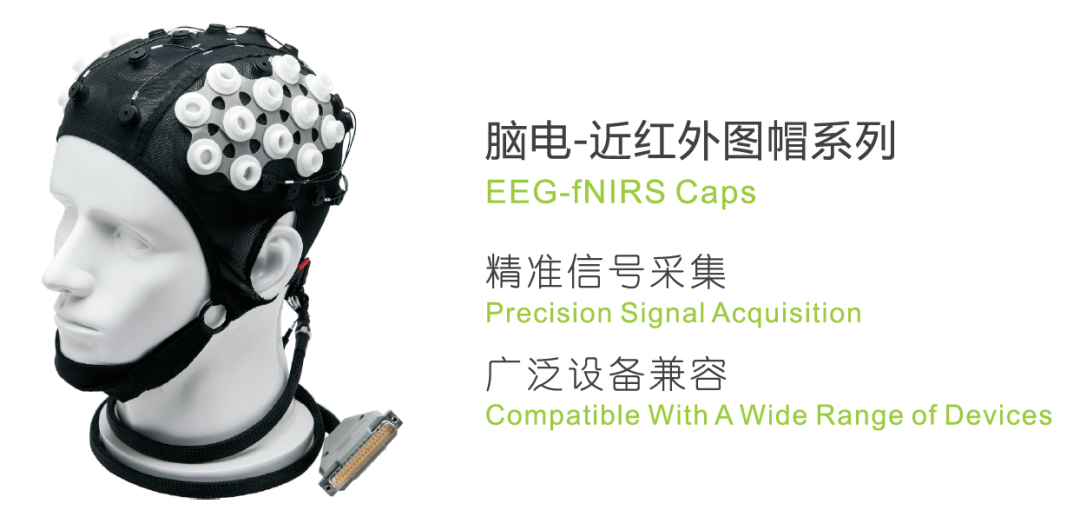
This study used the Green Tech EEG/fNIRS combined electrode cap,
Welcome to inquire for cooperation.
———–End of Sharing———–
Editor: Yezi
Reviewer: mingzlee7


Disclaimer: Some articles and information are sourced from the internet and do not represent the views of this subscription account or its authenticity. If the content involves copyright issues, please contact the editor Yezi immediately (WeChat ID: 1791438763), and we will take appropriate measures promptly. This subscription account’s original content requires authorization for reprinting, and the author and source must be indicated.