Text reproduced from Mudanjiang Tumor Hospital
Diffusion weighted imaging (DWI) is a technical method that utilizes the Brownian motion of water molecules to reflect the internal conditions of lesions, combined with MRI plain scans or enhancement sequences to achieve a clear diagnosis. The advantages include non-invasiveness, ease of operation, and a high detection rate for small lesions, allowing for the diagnosis of cerebral infarction within 30 minutes of onset, thus providing valuable time for thrombolytic therapy; it also has significant advantages in differentiating between benign and malignant tumors and assessing the treatment of malignant tumors. The Apparent Diffusion Coefficient (ADC) value, derived from the DWI sequence, is another reference indicator that can indirectly reflect lesion density. Extensive clinical research has shown its reference value for the benign or malignant nature of tumors and the specific staging of cerebral infarction. By combining the advantages of DWI and ADC characteristics, it is possible to obtain as much morphological and metabolic information about lesions as possible, aiding in the qualitative and staging of lesions, significantly improving diagnostic accuracy, and providing the most effective information for clinical decision-making regarding optimal treatment plans.
Specific clinical applications are as follows:
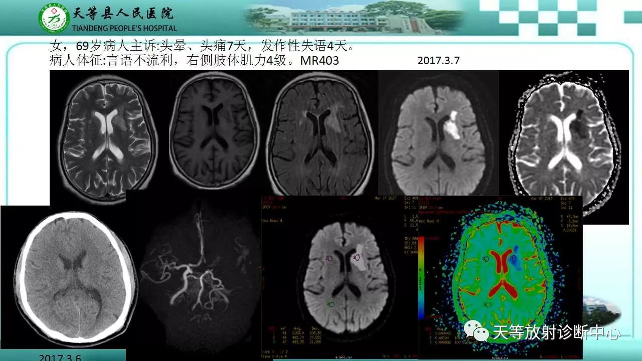
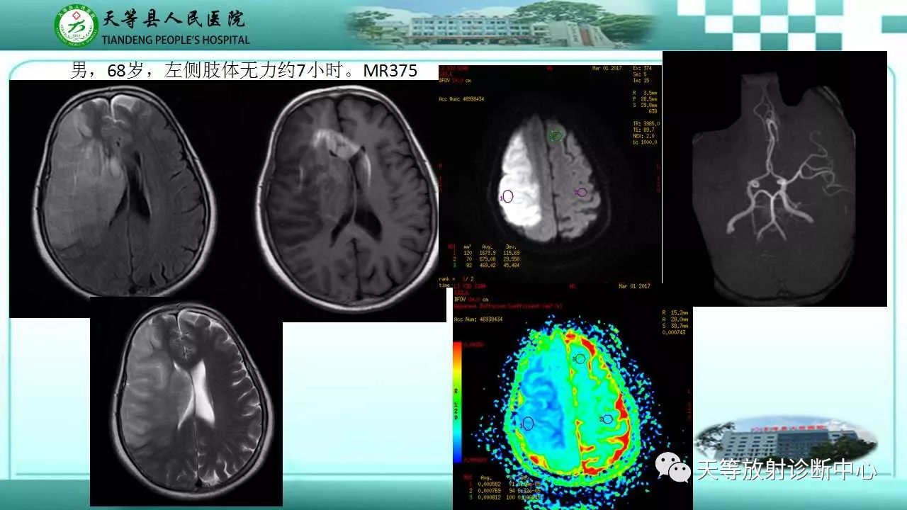
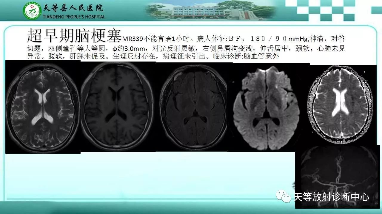
①Application in the nervous system: Most cerebral infarctions can be detected on DWI 30 minutes after the onset of infarction, showing restricted diffusion and reduced ADC values, reaching a minimum between 8 to 32 hours, and lasting for 3 to 5 days. In the acute phase, DWI shows high signals while the ADC map shows low signals.
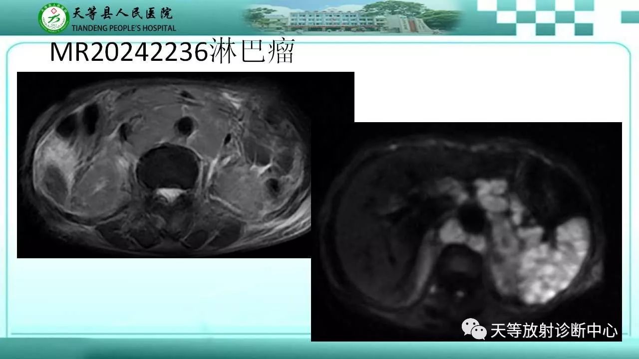
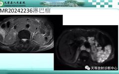
②Application in brain tumor diagnosis: The DWI characteristics of gliomas, metastatic tumors, and meningiomas vary. Some lesions present high signals on DWI, with ADC values showing either low or high signals. The size of the ADC value depends on the density of the tumor; high-grade gliomas have significantly lower ADC values than low-grade gliomas, primarily because higher malignancy in gliomas correlates with a greater number of cells and smaller intercellular spaces, as well as increased cellular atypia leading to more restricted water molecules. Conversely, a decrease in ADC values tends to indicate malignancy or atypical meningiomas, making DWI significant for differentiating between benign and malignant meningiomas. Additionally, lymphomas display significantly high signals on DWI with low ADC values due to their high tumor density, large nuclei, and low extracellular water content, allowing DWI to reliably differentiate lymphomas from other types of tumors when combined with other MRI sequences.
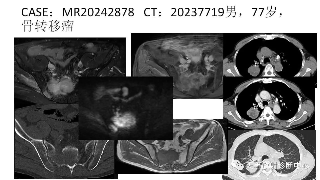
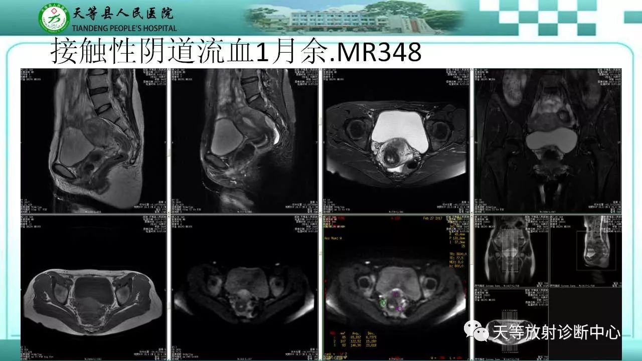
③Application in body tumor diagnosis: a. Malignant tumors usually present restricted high signals on DWI, with low ADC values. This application is widely used for the diagnosis and treatment evaluation of benign and malignant lesions in organs such as the liver, pancreas, uterus, appendages, gastrointestinal tract, bones, and breasts. For patients after chemotherapy, the reduction of restricted high signals on DWI and the increase of ADC values can indicate effective treatment; conversely, ineffective treatment is indicated by the opposite. b. Inflammatory lesions, tumor-like lesions, and benign tumors may show slightly restricted high signals or unrestricted low signals on DWI, with ADC maps showing equal or high signals and higher values. For example, focal nodular hyperplasia, hepatic adenoma, breast adenoma, and fibromas. A few exceptions exist, such as abscesses, where the abscess cavity on DWI shows restricted high signals, and ADC shows equal or low signals with low values, which can be differentiated by enhancement methods. Other inflammatory lesions or tumor-like lesions may show slight restriction or no restriction, with slightly high signals or equal/low signals, and higher ADC values.
④Application in prostate tumor diagnosis: DWI aids in the diagnosis, staging, differential diagnosis, and efficacy evaluation of prostate cancer. Most prostate cancer lesions show high signals on DWI, while prostatitis typically shows equal signals; the mean ADC value of prostate cancer is higher than that of prostatitis. Additionally, metastatic lymph nodes and bone metastases from prostate cancer also display significantly high signals on DWI.
⑤Whole body DWI technology: Whole body DWI requires whole-body or full-length cross-sectional scanning, three-dimensional reconstruction of images, ultimately forming PET-like images. Whole body DWI assists in the discovery of systemic metastases in advanced malignant tumors; it can also be used for the efficacy evaluation of hematological malignancies and systemic metastases.
As mentioned above, the application advantages of DWI technology and ADC values in MRI are increasingly prominent in the diagnosis and treatment of cerebrovascular diseases and tumor lesions.