Ophthalmic artificial intelligence, as an emerging element in the field of ophthalmology, has developed rapidly in the screening and diagnosis of eye diseases. In recent years, there has been in-depth research on common diseases such as cataracts and glaucoma, demonstrating significant application advantages and prospects, with some technologies already applied in clinical settings.
The “Science and Technology Bulletin” new media platform invited Professor Lin Haotian’s team from the Sun Yat-sen Ophthalmic Center to provide an in-depth interpretation of a study published earlier this year in the top cardiovascular journal Circulation, helping us understand the application value of ophthalmic artificial intelligence.
At the beginning of this year, Pradeep Natarajan’s team from Massachusetts General Hospital at Harvard Medical School published a research paper titled “Deep Learning of the Retina Enables Phenome- and Genome-Wide Analyses of the Microvasculature” in the top cardiovascular journal Circulation.
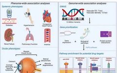
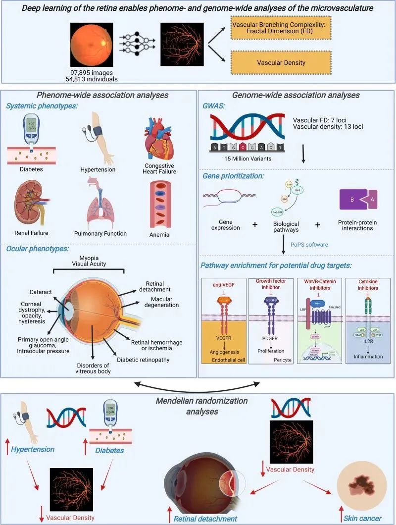
This study utilized Convolutional Neural Networks (CNN) to automatically segment the retinal microvascular structure and calculate vascular density (VD) and vascular fractal dimension (FD). Through phenotype-wide association studies (PheWAS) and genome-wide association studies (GWAS), it further explored the relationship between VD and FD and 1866 diseases based on the International Classification of Diseases, showcasing the tremendous potential of retina-based deep learning in understanding the human microvascular system and its future applications in screening and predicting various diseases.
Retinal vessels and their association with systemic phenotypes and genotypes
The Retina: A Window to the Systemic Microvascular System
The microvascular system affects the health of the whole body through different organs, and many ocular and systemic diseases are related to the dysfunction of the microvascular system.
Vascular development involves the differentiation of endothelial cells from mesodermal precursors and is regulated by various signaling pathways, including the WNT/β-catenin pathway and the Notch signaling pathway.
Vascular formation follows a series of orderly steps and is regulated by various growth factors and cytokines, including vascular endothelial growth factor (VEGF), fibroblast growth factor, angiopoietin, and intracellular signaling pathways involving Rho GTPases, protein kinase C, and Notch signaling.

The microvascular system also plays a significant role in the development of cancer, as neovascularization is essential for tumor growth and metastasis.
Anti-angiogenic drugs, such as anti-VEGF antibodies, are important means of cancer treatment; they are also the primary treatment methods for common ophthalmic diseases, such as wet age-related macular degeneration (w-AMD) and diabetic macular edema (DME).

Clearly, the microvascular system plays a critical role in maintaining normal organ function and tumor formation.
Given the anatomical and physiological similarities between retinal microvasculature and other organs, and the ability to achieve non-invasive in vivo assessment of retinal microvasculature through fundus photography, it serves as the best “window” for non-invasive assessment of the human microvascular system.
Deep Learning and Big Data:
A Powerful Tool for Ocular and Systemic Association Analysis
Data Collection and Quality Control
The UK Biobank is a long-term large-sample cohort that includes approximately 500,000 participants recruited from 2006 to 2010 (with a median follow-up time of 10 years), collecting and recording genomic and longitudinal phenotype data of the subjects.
The study first screened 97,895 fundus images from 67,339 participants in the above database, applying a quality control filter (a convolutional neural network developed by the team for automatic outlier detection), excluding 26% of the original images of poor quality, and ultimately included 97,895 fundus images from 54,813 participants for subsequent analysis research.
Retinal Vessel Segmentation and Feature Extraction
To achieve large-scale vascular segmentation, the study developed a U-Nets deep learning ensemble model on the Google Cloud AI platform. This model was developed based on 90 fundus photographs and corresponding hand-drawn retinal vascular segmentation images from three public datasets, to automate the segmentation of the retinal vascular system from fundus photographs.
Subsequently, U-Nets underwent internal and external validation, with results suggesting that compared to other previous convolutional neural networks, this model demonstrated comparable orbetter performance.
In an internal validation dataset containing 15 images, the Dice similarity coefficient (a measure of spatial overlap accuracy) of this ensemble model was 82.1%, pixel accuracy was 97.4%, and the area under the ROC curve was 99.1%; in an external validation dataset containing 143 images, the correlation of VD and FD with manually labeled true vascular parameters was 0.88 and 0.92, respectively.
PheWAS (Phenotype-Wide Association Study)
The study conducted four groups of PheWAS analyses corresponding to retinal VD and FD with: pre-existing phenotypes at enrollment; incident event phenotypes after enrollment; quantitative systemic biomarkers; and quantitative ocular traits.
Through PheWAS, the authors found that microvascular changes detected in the retina were associated with various diseases, providing evidence for the microvascular system as a biomarker to predict and assess the risk and severity of systemic diseases.
GWAS (Genome-Wide Association Study)
Using the Hail-0.2 software provided by Google Cloud for GWAS analysis of patients who had fundus retinal images collected at enrollment, adjusting for age, age squared, sex, smoking status (current/former/never), the top 10 principal components of genetic ancestry, and genotyping, linear regression models were applied for analysis.
Subsequently, polygenic risk score (PRS) PheWAS analysis, Mendelian randomization, and rare variant burden analysis were conducted.
Research Results
PheWAS
Association of Retinal Microvascular Changes with Systemic Diseases
Compared to patients without hypertension or diabetes, those with hypertension or diabetes and lower VD or PD had an increased risk of event mortality.
This indicates that lower microvascular density and branch complexity suggest that patients may have more severe cardiac metabolic diseases.
Additionally, the authors observed that lower retinal VD and FD were significantly associated with the prevalence and increased risk of the following diseases: cardiovascular metabolic diseases (such as increased body mass index, hypertensive heart disease, renal failure), pulmonary function abnormalities (such as sleep apnea), and hematologic system abnormalities (such as anemia).
Association of Retinal Microvascular Changes with Ocular Diseases
Although previous cohort studies have found a connection between retinal vascular-related indicators and diabetic retinopathy (DR), this study is the first large-scale study to determine the correlation between retinal vascular-related indicators and the occurrence of various ocular diseases.
Specifically, this study identified low VD and FD with multiple associations with posterior segment diseases (including retinal detachment, DR, macular degeneration, vitreous hemorrhage).
Notably, retinal detachment (RD) is a potentially blinding ocular disease, but currently identified risk factors associated with it are very limited, including ocular trauma, myopia, and family history.
The study found a significant correlation between lower retinal VD and FD and higher incidence of retinal detachment, and that both increase the risk of RD independently of myopia.
This suggests that early identification of individuals with low retinal vascular density can adjust the follow-up strategies for this high-risk population to further reduce their risk of blindness.

Furthermore, this study also identified the association of retinal VD and FD with anterior segment diseases (glaucoma, cataract), indicating that the retinal vascular system may also have physiological significance for other intraocular structures beyond the retina and vitreous.
The authors’ findings are consistent with the previously proposed hypothesis of a connection between the retinal vascular system and normal-tension glaucoma, which posits that the pathogenesis of normal-tension glaucoma is due to vascular abnormalities that hinder the delivery of nutrients to the retina, leading to degeneration of ganglion cells.

In summary, the above findings link the retinal vascular system with ocular pathophysiological changes, emphasizing the importance of the retinal microvascular system in maintaining ocular health, and helping us understand the relationship between retinal microvasculature and vision impairment through different mechanisms.
Data Collection and Quality Control
Genome-wide association analysis identified the connections between the retinal microvascular system and genes involved in angiogenesis, cancer, pigmentation, inflammation, and microvascular structure.
Genetic loci of retinal vascular-related indicators may provide therapeutic targets with pleiotropic effects for retinal lesions, cancer, and microvascular diseases in other tissues.
Angiogenesis
The study observed significant enrichment of pathways related to angiogenesis (VEGF, platelet-derived growth factor, angiopoietin), which are currently used in targeted therapies to inhibit neovascularization in DR, w-AMD, and many cancers.
Pigmentation
The study also observed a significant genetic correlation between the retinal vascular system and skin color, further fine-mapping significant genome-wide loci to prioritize potential causal variants.
On the fine-mapped variants, alleles that lower retinal VD and FD have heterogeneous effects on phenotypes previously assessed in genetic correlation analysis.
Specifically, this includes the following genes with high polygenic priority scores (PoPS): IRF4, SLC45A2, OCA2, DUSP22, ACTG1.
OCA2 encodes the oculocutaneous albinism type 2 protein, known to lead to lighter skin color, increasing susceptibility to skin cancer.
Additionally, harmful variants of the MITF (microphthalmia-associated transcription factor), essential for normal melanocyte differentiation, were also discovered in rare variant association studies and GWASs, which are associated with Waardenburg syndrome, characterized by abnormal pigmentation of the eyes, hair, and skin.
Previous studies have also found that MITF protein labeling in human tumor samples is significantly enhanced around blood vessels.
Inflammatory Response
In two GWAS studies, inflammatory and chemokine pathways were significantly enriched, with contributing genes prioritized in PoPS including IL2RA, IL23A, IL1R2, IL2, IL7R, IL6, MEF2A, PDGFRA, and HLA markers.
Interleukins (IL) are known to be involved in regulating angiogenesis in tumor formation, promoting or inhibiting blood vessel formation, and can inhibit tumor VEGF expression by suppressing IL.
Microvascular Structure
Two GWAS loci identified in the VD and FD genome-wide association analysis (GNB3 and GNB2) are key regulators of the chemokine signaling pathway, belonging to the G protein superfamily.
Notably, the missense variant Rs5442-A in GNB3 has previously been found to be associated with retinal microvascular diameter, hypertension, refractive errors, and age-related macular degeneration.
Single-Sample Mendelian Randomization
Mendelian randomization (MR) is a widely used experimental design method in epidemiology. It analyzes the causal relationships between exposure factors and outcomes by introducing a so-called “instrumental variable” as an intermediary variable.
This study found through MR analysis that individuals with genetic susceptibility to hypertension had lower retinal VD and FD.
Similarly, individuals with genetic susceptibility to type 2 diabetes also had lower retinal VD, but no significant association with FD was found, indicating that the correlation between diabetes and the retinal microvascular system may stem from vascular diameter rather than complexity of branching, consistent with previous conclusions regarding retinal vascular diameter.

Furthermore, individuals with a congenital low vascular density genotype had a higher risk of myopia and retinal detachment (independent of myopia and equivalent spherical power), as well as a higher risk of skin cancer (independent of major genetic factors, skin color, sun exposure, and photosensitivity).
Notably, the genetic link between retinal vascular density and retinal detachment, as well as the phenotypic link between VD and RD events, emphasizes the potential causal relationship between the two, potentially identifying a new risk factor for retinal detachment that could be applied to future monitoring and treatment of RD.
Retina-Based
Systemic Disease Assessment May Become Possible
This study utilized a deep learning ensemble model to quantitatively analyze nearly 100,000 fundus photographs, using VD and PD to identify clinical associations and genomic risk factors of the retinal microvascular system with the full phenotype.
From an epidemiological perspective, low VD and PD are associated with increased risk of long-term event mortality, cardiovascular metabolic diseases, and ocular disease occurrence; from a genetic perspective, VD and PD are significantly enriched in pathways related to angiogenesis and inflammation.
The above research results demonstrate the tremendous potential of retina-based deep learning in understanding the human microvascular system and the possibility of its future broad applications in screening and predicting various diseases.
Retinal Microvascular Index could serve as a biomarker for the severity of cardiovascular metabolic diseases, potentially enabling early identification of high-risk populations for cardiovascular event mortality (such as arrhythmia, congestive heart failure, etc.) in the future, reducing their long-term mortality rate.

Similarly, previous studies have utilized deep learning models to establish qualitative associations between ocular features (conjunctiva, sclera, iris, and fundus) and major hepatobiliary diseases (liver cancer, cirrhosis, chronic viral hepatitis, non-alcoholic fatty liver, cholelithiasis, liver cysts), achieving automatic screening and identification of common hepatobiliary diseases through ocular images.
Particularly in identifying liver cancer and cirrhosis, the model demonstrated good diagnostic performance (AUC reaching 0.90).
In the future, this AI model may provide a simple, rapid, non-invasive new method for large-scale screening of hepatobiliary diseases.

The study also has the following limitations:
First, factors that reduce image quality (such as cataracts and fundus pigmentation) may affect and confuse the associations between phenotype and genotype. However, after adjusting for cataracts, retinal detachment, myopia, and skin color, the associations between retinal vascular features and systemic characteristics remained largely unchanged.
Second, although TOPCON images are generally uniform in magnification, the magnification of images is related to individuals’ equivalent spherical power. However, after sensitivity analysis adjusted for equivalent spherical power and myopia, the associations remained consistent.
Third, the current analysis is based on the UK Biobank, which is primarily composed of Europeans and has only limited fundus images obtained from TOPCON OCT scanners. Future studies in differently racial populations and other imaging modalities are necessary.

In conclusion, the author believes that artificial intelligence has unique advantages and tremendous potential in analyzing vast amounts of data and images, far exceeding human computational capabilities. It may help explore and discover subtle biological events and their intrinsic connections that have not yet been observed by humans, providing a new strategy and method for interdisciplinary disease research and automatic screening.
References:
[1] ZEKAVAT S M, RAGHU V K, TRINDER M, et al. Deep Learning of the Retina Enables Phenome- and Genome-Wide Analyses of the Microvasculature [J]. Circulation, 2022, 145(2): 134-50.
[2] XIAO W, HUANG X, WANG J H, et al. Screening and identifying hepatobiliary diseases through deep learning using ocular images: a prospective, multicentre study [J]. Lancet Digit Health, 2021, 3(2): e88-e97
Authors: Li You, Cui Tingxin
Review Expert: Lin Haotian, Deputy Director of the Sun Yat-sen University Sun Yat-sen Ophthalmic Center, Chief Physician, Professor, Researcher, Dual-disciplinary Doctoral Supervisor. Member of the National Youth Federation Standing Committee, Vice Chairman of the Guangdong Provincial Youth Federation, Director of the Asian Office of the Asia-Africa Ophthalmology Society. Chief Scientist of the National Key Research and Development Program, National “Ten Thousand Talents Plan” Leading Talent, Outstanding Contribution Young and Middle-aged Expert of the National Health Commission, recipient of the National May Fourth Youth Medal, Wu Wenjun Artificial Intelligence Science and Technology Progress First Prize, and the First Zhong Nanshan Youth Science and Technology Innovation Award. He has published nearly 200 high-level papers as the first or corresponding author (including co-authorship) in journals such as Science, Nature, Nature Biomedical Engineering (3 articles), and Lancet Digital Health (2 articles). His achievements were selected by IEEE Spectrum as one of the “11 AI Events Impacting Global Medicine” and recognized by the National Health Commission as one of the “Top Ten Medical Technology News in China in 2019.” He has been granted (applied for) 26 invention or utility patents and 21 software copyrights, and has completed the transformation and application of 4 intelligent technology devices.
Highlights:
·The Eighth Beijing-Tianjin-Hebei Dry Eye Diagnosis and Treatment Seminar Concluded Successfully (Includes Replay Link)
·New Minimally Invasive Surgical Treatments for Angle-Closure Glaucoma
·[Insights] Professor Zeng Qingyan: Dry Eye Diagnosis and Treatment from the Perspective of Socioeconomics
·Exquisite Work丨New Book Release of “Expert Techniques in Ophthalmic Surgery (2nd Edition)”
·Wang Ningli, Sun Xinghuai, and Zhang Xiulan Selected Among the Top 30 Global Glaucoma Experts
·“The Seventh Dry Eye Conference” Grandly Opened Online! Domestic and foreign experts gathered to discuss dry eye
·Collaborative Innovation for Development—The Seventh National Dry Eye Academic Conference, Fifth National Conference on Ocular Surface and Tear Liquid Diseases, and Fifth National Conference on Traditional Chinese Medicine Eye Care Concluded Successfully
·“The Seventh Dry Eye Conference” Grandly Opened Online! Domestic and foreign experts gathered to discuss dry eye
·2020 China Dry Eye Expert Consensus Interview with Professor Liu Guoguang—Based on Practice, Integrating Innovation, Building Consensus, and Composing a New Chapter in Dry Eye Development
·China’s Consensus on the Diagnosis and Treatment of New Vascular Glaucoma (2019)
·Public Lectures, Literature, Experts Lead You to Fully Interpret Hot Issues in Dry Eye