Journal of Practical Neurology in China
For learning and communication only, please indicate the source when reprinting!
Claude syndrome was first described in 1912 by French neurologist Claude, also known as red nucleus syndrome. The main symptoms include ipsilateral oculomotor nerve palsy and contralateral ataxia. Oculomotor nerve palsy may involve pupillary dilation and limited eye movement, with anatomical localization described as the red nucleus, oculomotor nerve bundle, and superior cerebellar peduncle. This syndrome results from a specific infarction in the midbrain, which can lead to delayed treatment. Here, we report a case of Claude syndrome admitted to our hospital.
1. Clinical Data
The patient, a 55-year-old female, was admitted with the main complaint of “double vision and unsteady walking for 6 days, left eyelid ptosis for 5 days”.Six days prior, the patient experienced double vision at rest without obvious cause, which became more pronounced when looking to the left, along with unsteady walking, needing support while walking, and right-hand weakness. There was no significant dizziness, visual rotation, limb numbness, or seizures, and no speech impairment. Bowel and bladder function were normal. On the second day of illness, she went to a local county hospital, where a CT scan of the head and cardiac ultrasound showed no significant abnormalities. One day later, left eyelid ptosis appeared, and the local hospital initially diagnosed “oculomotor nerve palsy” and provided unspecified treatment, but her condition did not improve, leading to her transfer to our hospital on the sixth day of illness.The patient had a 10-year history of hypertension, taking 1 tablet of nifedipine per day, and a past history of “stroke”, denying diabetes or heart disease. Upon admission, examination revealed:Blood pressure 122/77 mmHg, alert and articulate, normal cognitive function, left eyelid ptosis, left pupil 4 mm with sluggish light reflex, right pupil 3 mm with sensitive light reflex, limited adduction, upward, and downward movement of the left eye, unlimited movement of the right eye in all directions, normal muscle strength and tone in limbs, symmetrical bilateral limb and facial sensation, and negative bilateral pathological signs;Right finger-to-nose test and heel-to-knee test were inaccurate, and Romberg’s sign was positive;Meningeal irritation signs were negative.Auxiliary examinations showed:Triglycerides 3.36 mmol/L, homocysteine 16 mmol/L, with other biochemical indicators and blood routine basically normal.Cervical vascular ultrasound indicated:Bilateral carotid intima-media thickening with plaque formation; after admission,head MRI + DWI + MRA (Figure 1):(1) New cerebral infarction in the midbrain;Bilateral basal ganglia, bilateral thalamus, and brainstem hemorrhagic sequelae;(2) Bilateral periventricular white matter demyelination changes;(3) MRA of the brain indicated left middle cerebral artery M1 segment stenosis with reduced distal branches;Bilateral posterior cerebral arteries were thin and had reduced signals.After admission, treatment included antiplatelet aggregation, plaque stabilization, circulation improvement, oxygen free radical clearance, blood pressure regulation, and blood lipid adjustment.After 10 days of treatment, the patient’s eye movement improved compared to before, but had not returned to normal levels. The left eyelid ptosis slightly improved, and right-sided ataxia showed improvement, enabling her to eat and walk independently, and she was discharged after improvement.
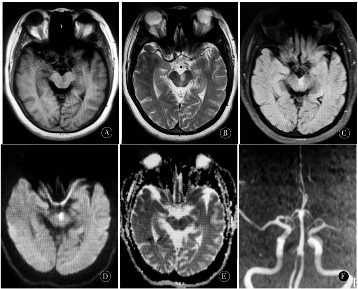
Figure 1 A, B, C: Left midbrain medial long T1 long T2 high FLAIR abnormal signal; D: DWI shows high signal on the left midbrain; E: ADC shows low signal on the left midbrain; F: MRA shows left middle cerebral artery M1 segment stenosis with reduced distal branches; bilateral posterior cerebral arteries were thin and had reduced signals (head MRI + DWI + MRA on the 7th day of onset)
2. Discussion
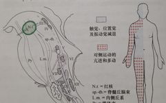
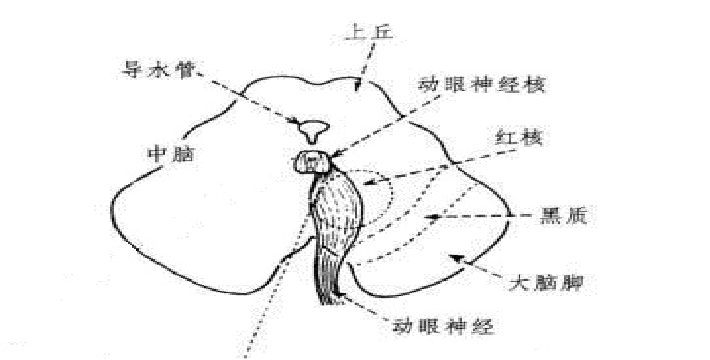
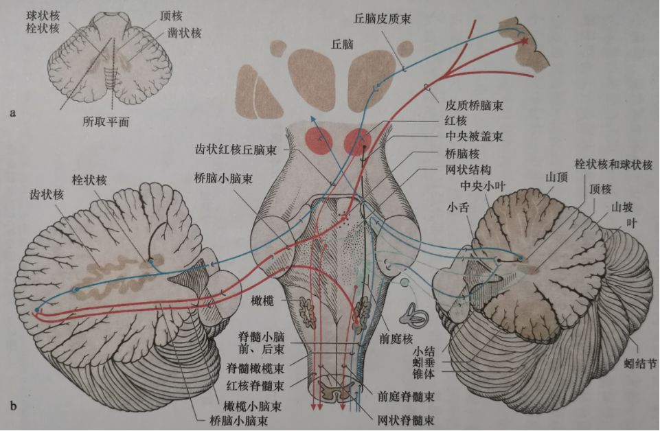
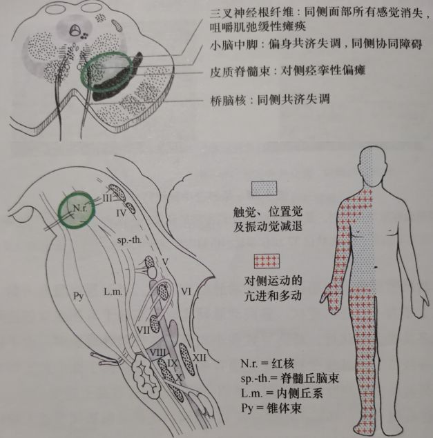
The oculomotor nuclei located in the midbrain tegmentum are distributed relatively dispersedly. The root fibers of these nuclei travel ventrally along with parasympathetic fibers, partially traversing the red nucleus, ultimately forming the oculomotor nerve that exits the brainstem bilaterally from the interpeduncular fossa.The afferent impulses to the red nucleus come from the cerebellar emboliform and dentate nuclei. The cerebellar-red nucleus bundle crosses within the superior cerebellar peduncle in the midbrain, participating in the regulation of body posture and assisting in the precise and smooth execution of voluntary movements (as shown in the schematic diagram below).When near the aqueduct of Sylvius at the level of the superior colliculus, tumors, inflammation, and vascular lesions can affect the ipsilateral oculomotor nerve fiber bundle and the outgoing fibers of the superior cerebellar peduncle, resulting in a group of symptoms including ipsilateral oculomotor nerve palsy and contralateral cerebellar ataxia.
This syndrome is rare, and since its first report, there have been only a few case reports domestically and internationally, with differing opinions on the localization of the responsible lesion. KREMER initially described Claude syndrome as involving the red nucleus.DHANJAL et al. conducted pathological examinations on a case of Claude syndrome, revealing a pericentral midbrain infarction affecting part of the superior cerebellar peduncle and the red nucleus, while MRI showed no lesions in the red nucleus, indicating that Claude syndrome does not necessarily involve the red nucleus. However, it should be noted that MRI findings may not fully align with the areas affected by neuropathology.SEO et al. studied six cases of Claude syndrome and CHIN-SHIH et al. conducted imaging and pathological research on one case, finding that the anatomical localization of this syndrome primarily involves the superior cerebellar peduncle in addition to the oculomotor nerve bundle, with the red nucleus only mildly affected.The paramedian artery of the midbrain is one of several branches of the posterior cerebral artery, supplying the paramedian zone of the midbrain, including the medial parts of the substantia nigra, red nucleus, and superior cerebellar peduncle, as well as the oculomotor nucleus and nerve roots.The paramedian artery of the midbrain is a small terminal artery, and these branches are susceptible to obstruction by microatheromatous emboli or cardiac emboli, which can also lead to hypoperfusion ischemic damage. Stenosis of the posterior cerebral artery is considered a potential factor in these patients.In conventional vascular imaging such as head MRA and CTA, it is often difficult to detect this artery. DSA can clearly identify the presence of this artery, but it is an invasive examination and not a routine check, so it is often inferred based on the ischemic area.The patient’s head MRA showed left middle cerebral artery M1 segment stenosis with reduced distal branches;Bilateral posterior cerebral arteries were thin and had reduced signals.The patient’s cardiac ultrasound showed no abnormalities, suggesting that the onset was related to atherosclerotic thrombosis or arterial stenosis of the midbrain supply arteries due to hypertension. Pure midbrain infarction accounts for 0.6% of ischemic strokes, and ataxia is the most common clinical manifestation of midbrain infarction, likely due to the presence of rich nerve fiber bundles connecting to the cerebellum. Oculomotor nerve palsy has been considered a clinical feature of midbrain infarction, but occurs in only 35% of pure midbrain infarctions, and patients with simultaneous pupillary dilation are less frequently observed, indicating that a small portion affects all oculomotor nuclei and nerve bundles. Therefore, sudden onset of oculomotor nerve palsy and ataxia should be taken seriously to rule out the possibility of stroke.The patient suddenly developed left eyelid ptosis, left eye adduction, and inability to look up and down, with pupillary dilation, indicating complete oculomotor nerve palsy. The oculomotor nerve bundle was damaged, and right-sided ataxia was present, causing her to easily tilt to the right, indicative of damage to the cerebellar connecting fibers, possibly affecting the cerebellar red nucleus and the red nucleus;On the 7th day of illness, the patient underwent head MRI + DWI + MRA, indicating a new lesion in the left midbrain anterior medial region, with atherosclerotic basis in the carotid and cerebral arteries. Considering the patient’s middle-aged female status, acute onset, past history of hypertension and stroke, it can be clearly localized as midbrain infarction. In this case, there were no tremors, rigidity, or involuntary movements in the contralateral limbs, thus differing from Benedikt syndrome (red nucleus syndrome, as shown in the figure below), making it a more typical case of Claude syndrome.The patient visited the local county hospital on the second day of illness, underwent head MRI, but did not have DWI, and no obvious lesions were found. The grassroots hospital’s diagnosis was unclear, leading to delayed treatment.Benedikt syndrome (red nucleus syndrome)
Isolated oculomotor nerve palsy with pupillary dilation is most commonly caused by diabetes or microvascular disease leading to ischemia of cranial nerves. Most oculomotor nerve palsies are peripheral damage. For patients with risk factors for stroke such as hypertension, diabetes, and history of stroke, if sudden unilateral oculomotor nerve palsy occurs, careful examination for other neurological signs should be conducted. If accompanied by contralateral cerebellar ataxia, midbrain infarction should be considered, with the possibility of Claude syndrome. Timely head MRI examination, especially DWI, should be completed for early diagnosis and treatment to provide the best recovery for the patient.Authors: Peng Xiaoxue, Bao Sujuan, Ma Xiaojun, Ge Zhaoming Long press the QR code to follow us
Neurology Medical Network
Helping Physicians Grow