
Iodinated contrast agents are widely used in CT angiography (CTA) examinations and play an irreplaceable role in the imaging diagnosis of vascular diseases throughout the body. However, there are risks of adverse reactions to injected contrast agents, contraindications, and high costs.
Recently, a new study conducted by the First Medical Center of the PLA General Hospital Radiology Department team led by Professor Lou Xin and Professor Fu Ying’s team from Beijing Institute of Technology has discovered:
It is possible to synthesize images comparable to real CTA quality without using contrast agents. This research was published in the top radiology journal Radiology.
The team has developed a system that can “synthesize CTA images,” a CTA imaging model based on Generative Adversarial Networks (CTA-GAN model), which can generate effects comparable to real CTA images. This system can assist in diagnosing aortic and carotid artery diseases while avoiding the risks associated with contrast agent use.
● Article published in Radiology

Non-Contrast Enhanced CT Angiography of Aorta and Carotid Arteries Based on Generative Adversarial Networks (GAN)
Establish a deep learning imaging model that does not require the injection of contrast agents for synthesizing CTA images, and evaluate the quantitative and qualitative image quality and diagnostic accuracy of synthesized CTA (Syn-CTA) images.
The Generative Adversarial Network-based CTA imaging model (CTA-GAN model) retrospectively collected paired NCCT-CTA (non-contrast-enhanced CT and CT angiography) data from the neck and abdomen over nearly 6 years for training, validation, and testing, and further validation on external datasets.
From January 2017 to December 2022, paired NCCT-CTA data from the neck and abdomen were collected at the First Medical Center of the PLA General Hospital, which serves as the internal test set. Additionally, paired abdominal NCCT-CTA data collected from June 2018 to March 2023 at Hunan Brain Hospital serves as the external test set.
The internal test set collected data from 1,749 patients (median age: 60 years; IQR: 50–68 years; male: 1057) with neck and abdominal CT images. Among them, images from 1,137 patients were used to train the model, 400 for validation, and 212 for testing. The external validation set included CT images from 42 patients (median age: 67 years; IQR: 59–74 years; male: 37).
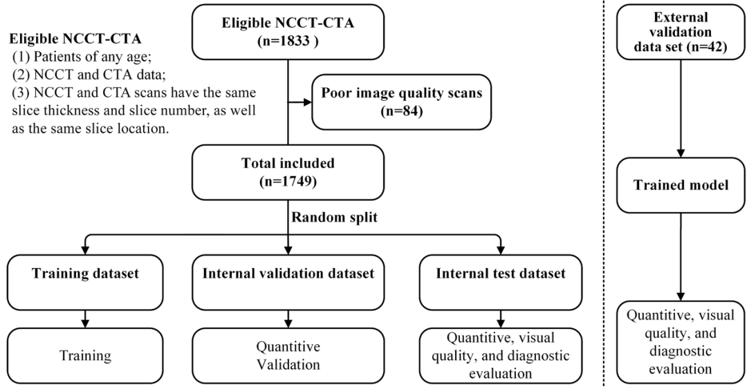
● Flowchart of paired NCCT and CTA scans included in the study
All CTA examinations used iodinated contrast agent, with a concentration of 370 mg/ml and an injection speed of 4.5 ml/sec.
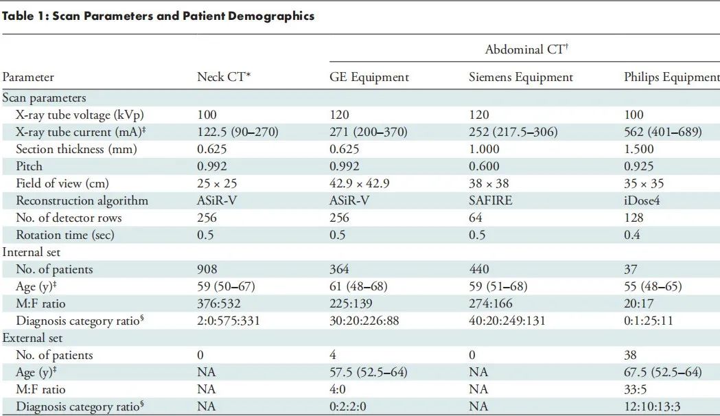
Figure shows patient scanning parameters and statistics, including:
Neck CT: Scanned using GE equipment, covering at least from the jugular bulb to the aortic arch.
Abdominal CT: Scanned covering at least the diaphragm of the iliac arteries; chest + abdominal CT angiography scans are included in this category.
Diagnosis Categories: Aneurysm, atherosclerosis, healthy arteries.
❶ Use quantitative metrics to evaluate the quality of “synthesized CTA” images.
❷ Two experienced radiologists performed a three-point scoring (3 = good) for visual quality and determined the diagnosis of vascular diseases.
❸ The effectiveness of synthesized CTA images is assessed by comparing visual quality scores and diagnostic accuracy for aortic and carotid artery diseases between synthesized CTA and real CTA scans.
❶ Synthesized CTA images are highly similar to real CTA images (normalized mean absolute error of 0.011 and 0.013 for internal and external test sets, respectively; peak signal-to-noise ratio of 32.07 dB and 31.58 dB; structural similarity of 0.919 and 0.906);
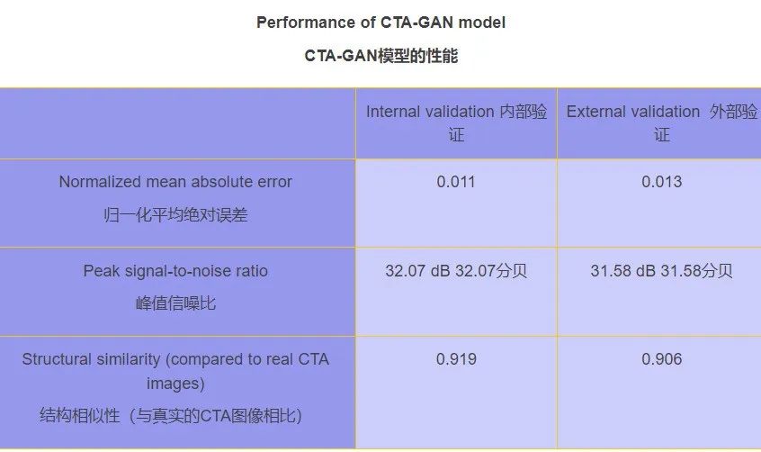
● Performance of the CTA-GAN model
❷ The visual quality of synthesized CTA images is comparable to real CTA images (internal test set, P = .35; external validation set, P > .99);
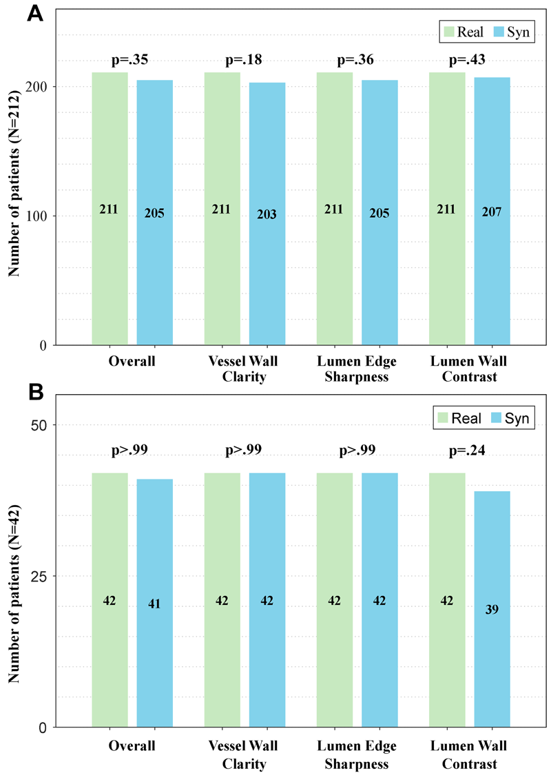
● Comparison of visual quality scores between synthesized (Syn) and real CT angiography (Real) images in (A) internal test set and (B) external validation set.
❸ Synthesized CTA shows reasonable to good diagnostic accuracy for vascular diseases (internal test set: accuracy = 94%, macro F1 score = 91%; external validation set: accuracy = 86%, macro F1 score = 83%).
CTA images of the neck or abdomen based on the CTA-GAN model do not require iodinated contrast agents, and the image quality is comparable to real CTA images, with reasonable to good diagnostic accuracy, showing broad application prospects in the diagnosis of vascular diseases.

Generative Adversarial Network-based CTA Imaging Model (CTA-GAN Model)
By normalizing the intensity within the range of -2000 to 2095 pixels to -1 to 1, each volume is processed independently. Based on inclusion and exclusion criteria, data with poor image quality were manually excluded. The final obtained data was randomly divided into training set, validation set, and test set (using scan as the basic unit) for model development.
CTA-GAN model includes the following components:
-
A CTA image synthesis generator based on CT plain scan images;
-
Corrector: Used to adjust CT images to account for error loss;
-
Discriminator: Used to estimate the probability that the input image is a real CTA image versus a synthesized image.
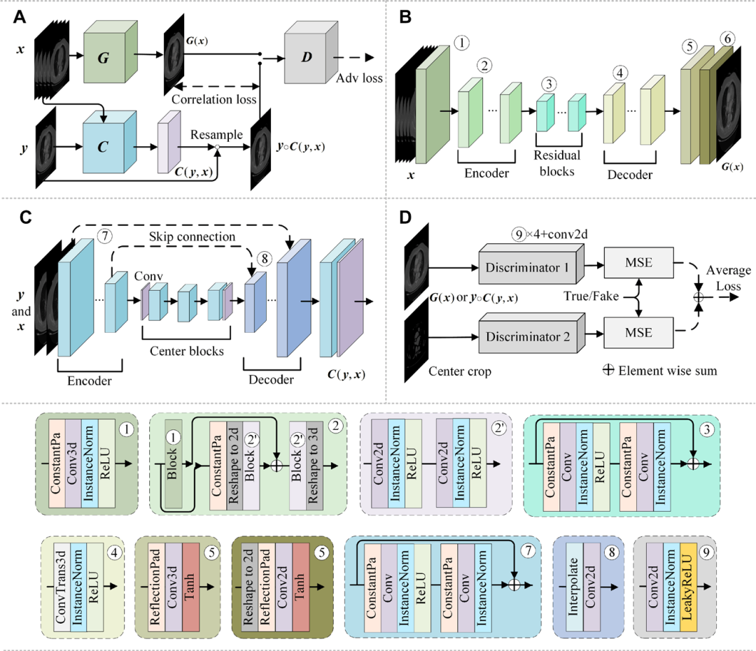
● Network architecture diagram of the CTA-GAN model

Quality of Synthesized CTA Images from Different Models
Let’s first look at the comparison of non-contrast-enhanced CT (NCCT) images of healthy patients, real CT angiography (CTA) images, and synthesized CTA images based on the Generative Adversarial Network (CTA-GAN) and two other models (pix2pix and RegGAN) in the internal test set.
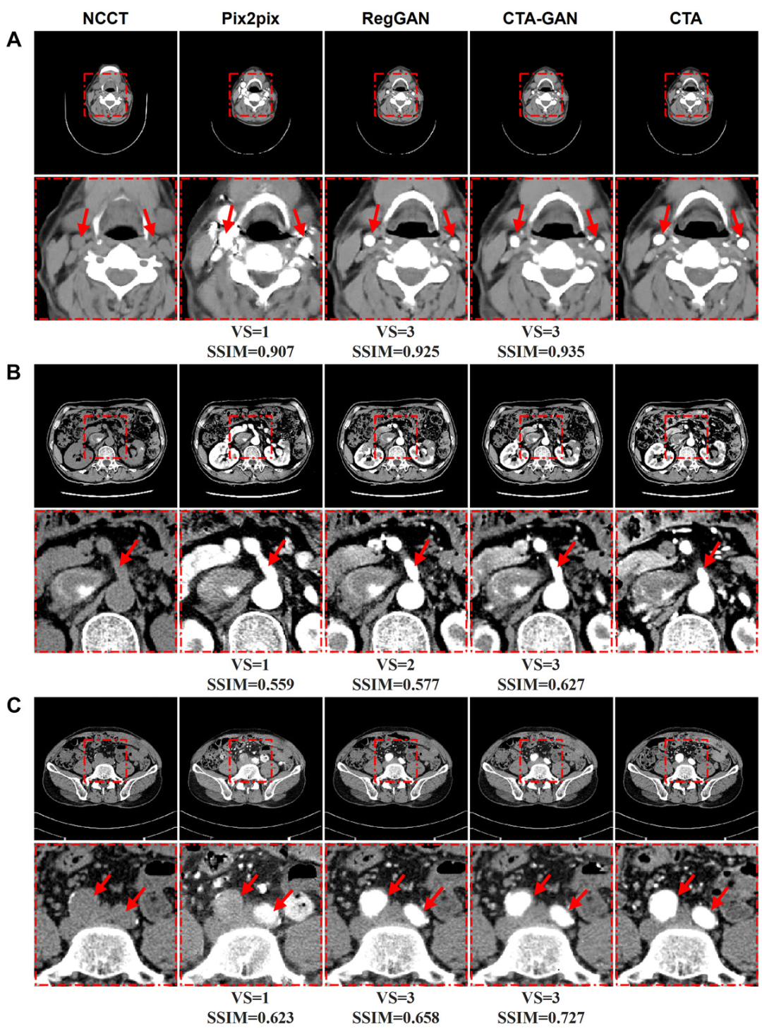
Each row of images represents the same axial plane, using real CTA images (right) as a reference. The area in the red box of the upper image of each example is enlarged and displayed below.
(A) Image of a 43-year-old female patient with carotid artery.
(B) Image of a 71-year-old male patient with abdominal aorta.
(C) Image of a 57-year-old male patient with abdominal aorta.
Arrows indicate target vascular structures. SSIM = Structural Similarity Index Measure, VS = Visual Score.
Now let’s look at the comparison of non-contrast-enhanced CT (NCCT) images of patients with vascular diseases, real CT angiography (CTA) images, and synthesized CTA images based on the Generative Adversarial Network (CTA-GAN) and two other models (pix2pix and RegGAN).
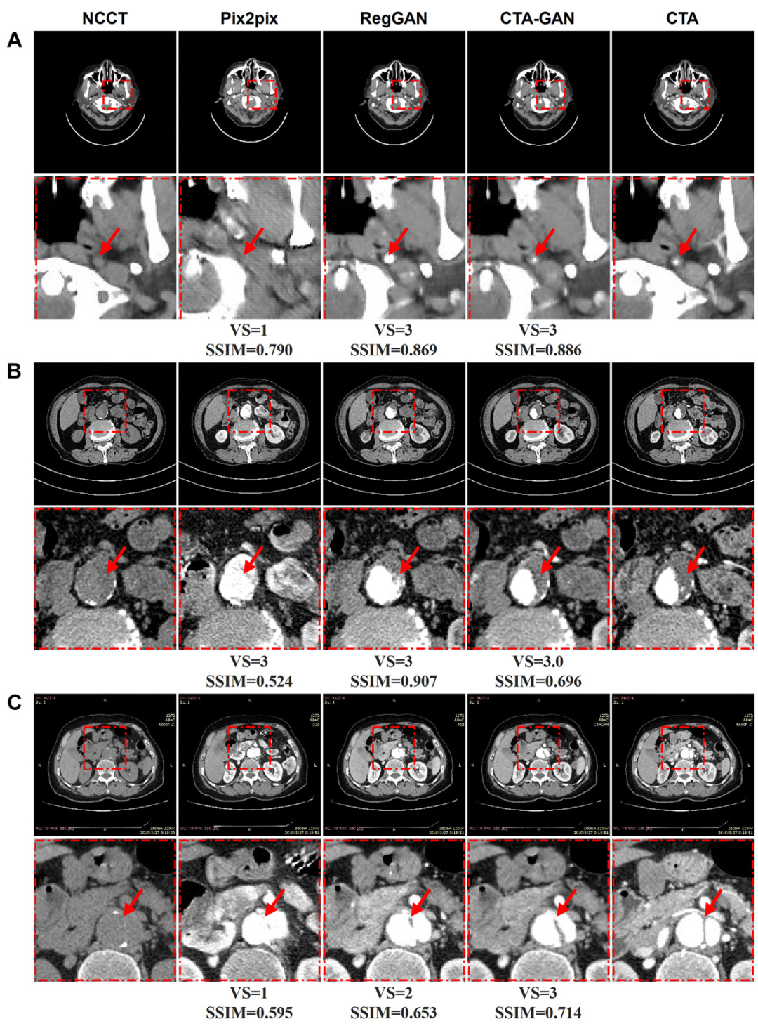
(A) Image of a 54-year-old male patient with internal carotid artery stenosis showing significant stenosis (arrow).
(B) Image of a 74-year-old male patient with abdominal aortic aneurysm showing aneurysmal dilation, with significant thickening of the abdominal aorta wall (arrow), indicating mural thrombus.
(C) Image of a 62-year-old male patient with abdominal aortic dissection showing no calcified intimal line, but with some calcification deposits in the vessel wall (arrow).
SSIM = Structural Similarity Index Measure, VS = Visual Score.

This research result has received significant attention
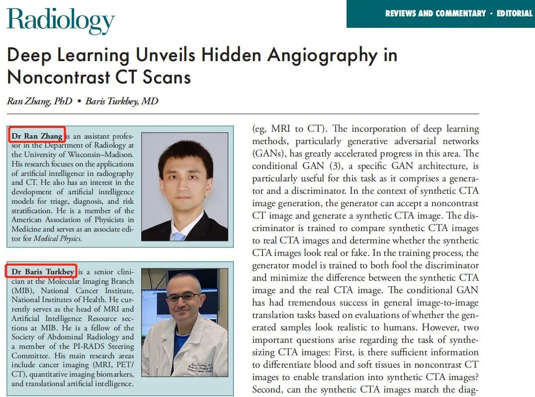
Professor Ran Zhang from the University of Wisconsin-Madison Radiology Department and Professor Baris Turkbey from the National Cancer Institute of the NIH published a commentary simultaneously, noting:
This work demonstrates the feasibility of generating CTA images from non-contrast-enhanced CT images using deep learning, laying the foundation for diagnosing major vascular diseases based on non-contrast-enhanced CT.
In other words, the development of the CTA-GAN model has created a non-contrast agent alternative for CTA, providing another option for those allergic to iodine and others with contraindications for contrast-enhanced examinations, and may also reduce the occurrence of adverse events related to contrast agents.
It is reported that there have been no prior reports on the generation of CTA images using plain CT images without any contrast agent for clinical diagnosis, so this technology has attracted close attention from the renowned medical AI company Terarecon in the United States, which immediately reported on the article online after its publication.
● Reports on this technology from foreign media
The successful development of this new technology
opens the era of non-contrast CT major vascular imaging
which will greatly assist in health check-ups and the construction of chest pain centers
Professor Lou Xin’s team’s research results are exciting
We feel very proud
Looking forward to further research in the future
bringing more good news